. 2023 Oct 18;24(20):15294.
doi: 10.3390/ijms242015294.

Effect of the Functional VP1 Unique Region of Human Parvovirus B19 in Causing Skin Fibrosis of Systemic Sclerosis

Affiliations

Effect of the Functional VP1 Unique Region of Human Parvovirus B19 in Causing Skin Fibrosis of Systemic Sclerosis

Der-Yuan Chen et al. Int J Mol Sci. .

Abstract

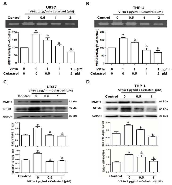
Human parvovirus B19 (B19V) is a single-stranded non-enveloped DNA virus of the family Parvoviridae that has been associated with various autoimmune disorders. Systemic sclerosis (SSc) is an autoimmune connective tissue disorder with high mortality and has been linked to B19V infection. However, the precise mechanism underlying the B19V contribution to the development of SSc remains uncertain. This study investigated the impacts of the functional B19V-VP1 unique region (VP1u) in macrophages and bleomycin (BLE)-induced SSc mice. Cell experimental data showed that significantly decreased viability and migration of both B19V-VP1u-treated U937 and THP-1 macrophages are detected in the presence of celastrol. Significantly increased MMP9 activity and elevated NF-kB, MMP9, IL-6, TNF-α, and IL-1β expressions were detected in both B19V-VP1u-treated U937 and THP-1 macrophages. Conversely, celastrol revealed an inhibitory effect on these molecules. Notably, celastrol intervened in this pathogenic process by suppressing the sPLA2 activity of B19V-VP1u and subsequently reducing the inflammatory response. Notably, the administration of B19V-VP1u exacerbated BLE-induced skin fibrosis in mice, with augmented expressions of TGF-β, IL-6, IL-17A, IL-18, and TNF-α, ultimately leading to α-SMA and collagen I deposits in the dermal regions of BLE-induced SSc mice. Altogether, this study sheds light on parvovirus B19 VP1u linked to scleroderma and aggravated dermal fibrosis.

Keywords: VP1 unique region (VP1u); fibrosis; human parvovirus B19 (B19); macrophages; systemic sclerosis (SSc).


Conflict of interest statement

The authors declare no conflict of interest.

Figures

Figure 1
Figure 1
Effects of B19V-VP1u on macrophage survival and migration. Cell viability of (A) U937 and (B) THP-1 macrophages in the presence of 1 μg/mL B19V-VP1u and different concentrations of celastrol. Cell migration percentage of (C) U937 and (D) THP-1 macrophages in the presence of 1 μg/mL B19V-VP1u and different concentrations of celastrol. Similar results were obtained in three repeated experiments. The symbols * and & indicate significant differences between the unstimulated control group (0 μM) and the cells treated with 1ug/mL B19V-VP1u alone, respectively.
Figure 2
Figure 2
Effects of B19V-VP1u on MMP-9 activity and NF-kB expression in macrophage. MMP-9 activity of (A) U937 and (B) THP-1 cells in the presence of 1 μg/mL B19V-VP1u and different concentrations of celastrol. Expressions of MMP-9 and NF-kB in (C) U937 and (D) THP-1 cells in the presence of 1 μg/mL B19V-VP1u and different concentrations of celastrol. Ratios of MMP-9 and NF-kB on GAPDH were shown in the lower panel. Similar results were obtained in three repeated experiments. The symbols * and & indicate significant differences between the unstimulated control group (0 μM) and the cells treated with 1 μg/mL B19V VP1u alone, respectively.
Figure 3
Figure 3
Effects of inflammatory cytokine expressions in B19V-VP1u-activated macrophages. The concentrations of (A,D) IL-6, (B,E) TNF-α, and (C,F) IL-1β in the medium of U937 and THP-1 cells treated with 1 μg/mL B19V-VP1u and different concentrations of celastrol. Similar results were obtained in three repeated experiments. The symbols * and & indicate significant differences between the unstimulated control group (0 μM) and the cells treated with 1 μg/mL B19V VP1u alone, respectively.
Figure 4
Figure 4
Effects of B19V-VP1u on a mouse model of bleomycin-induced systemic sclerosis (BLE-SSc). (A) Schematic diagram of BLE-SSc fibrosis mouse model. (B) H&E staining and Masson’s trichrome staining of a mice dermis section from different groups. (C) Quantitative representation. (D) Hydroxyproline contents of mice dermal thickness from different groups (n = 3). Scale bar: 100 μm. The symbols *, &, and # indicate significant differences compared to the control group, BLE group, and BLE + VP1u group, respectively.
Figure 5
Figure 5
Detection of TGF-β, α-SMA, and collagen I in the back skin of mice. Expressions of (A) TGF-β, (B) α-SMA, and (C) collagen in the back skin of mice from different groups. Scale bar: 100 μm. (D) Quantitative results of positive signals. The symbols *, &, and # indicate significant differences compared to the control group, BLE group, and BLE + VP1u group, respectively.
Figure 6
Figure 6
Detection of IL-6, IL-17A, IL-18, and TNF-α in the back skin of mice. Expressions of (A) IL-6, (B) IL-17A, (C) IL-18, and (D) TNF-α in the back skin of mice from different groups. Scale bar: 100 μm. (E) Quantitative results of positive signals. The symbols *, &, and # indicate significant differences compared to the control group, BLE group, and BLE + VP1u group, respectively.
Figure 7
Figure 7
Expression of fibrosis-related genes in the skin tissues of mice. Relative mRNA expression levels of (A) TGF-β, (B) MMP-9, and (C) TNF-α in skin tissues of mice from different groups. The symbols * and & indicate significant differences compared to the control group, BLE group or BLE + VP1u group, respectively.

References

    1. Qiu J., Söderlund-Venermo M., Young N.S. Human Parvoviruses. Clin. Microbiol. Rev. 2017;30:43–113. doi: 10.1128/CMR.00040-16. - DOI - PMC - PubMed
    1. Zakrzewska K., Corcioli F., Carlsen K.M., Giuggioli D., Fanci R., Rinieri A., Ferri C., Azzi A. Human parvovirus B19 (B19V) infection in systemic sclerosis patients. Intervirology. 2009;52:279–282. doi: 10.1159/000232945. - DOI - PubMed
    1. Lin K.H., You S.L., Chen C.J., Wang C.F., Yang C.S., Yamazaki S. Seroepidemiology of human parvovirus B19 in Taiwan. J. Med. Virol. 1999;57:169–173. doi: 10.1002/(SICI)1096-9071(199902)57:2<169::AID-JMV14>3.0.CO;2-1. - DOI - PubMed
    1. Török T.J. Parvovirus B19 and human disease. Adv. Intern. Med. 1992;37:431–455. - PubMed
    1. Lombardo E., Ramírez J.C., Garcia J., Almendral J.M. Complementary roles of multiple nuclear targeting signals in the capsid proteins of the parvovirus minute virus of mice during assembly and onset of infection. J. Virol. 2002;76:7049–7059. doi: 10.1128/JVI.76.14.7049-7059.2002. - DOI - PMC - PubMed