Review
. 2023 Oct 18;24(20):15301.
doi: 10.3390/ijms242015301.

Recent Advances in In Vivo Somatic Cell Gene Modification in Newborn Pups

Affiliations
Review

Recent Advances in In Vivo Somatic Cell Gene Modification in Newborn Pups

Shingo Nakamura et al. Int J Mol Sci. .

Abstract

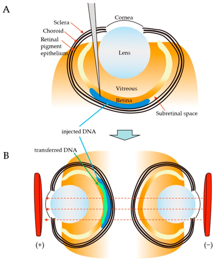
Germline manipulation at the zygote stage using the CRISPR/Cas9 system has been extensively employed for creating genetically modified animals and maintaining established lines. However, this approach requires a long and laborious task. Recently, many researchers have attempted to overcome these limitations by generating somatic mutations in the adult stage through tail vein injection or local administration of CRISPR reagents, as a new strategy called "in vivo somatic cell genome editing". This approach does not require manipulation of early embryos or strain maintenance, and it can test the results of genome editing in a short period. The newborn is an ideal stage to perform in vivo somatic cell genome editing because it is immune-privileged, easily accessible, and only a small amount of CRISPR reagents is required to achieve somatic cell genome editing throughout the entire body, owing to its small size. In this review, we summarize in vivo genome engineering strategies that have been successfully demonstrated in newborns. We also report successful in vivo genome editing through the neonatal introduction of genome editing reagents into various sites in newborns (as exemplified by intravenous injection via the facial vein), which will be helpful for creating models for genetic diseases or treating many genetic diseases.

Keywords: CRISPR/Cas9; genetically modified animal; genome editing; in vivo genome engineering; newborn; somatic mutation.


Conflict of interest statement

The founding sponsors had no role in the study design; the data collection, analyses, or interpretation; the writing of the manuscript; or the decision to publish the results.

Figures

Figure 1
Figure 1
Schematic representation of various routes of neonatal gene delivery: Newborn pups were subjected to intracerebral, intramuscular (i.m.), skin-targeted, or intraperitoneal (i.p.) injection. Solution injection is also possible intravenously (i.v. injection) via the retro-orbital sinus, temporal facial vein, or jugular vein. The figure was drawn in-house.
Figure 2
Figure 2
Schematic of in vivo gene delivery via subretinal injection into newborn mouse pups: (A) Subretinal injection. The blunt Hamilton needle was pushed through the retina as described by Venkatesh et al. [115]. The needle stops at the sclera if it is not pushed hard, because of the tougher composition of the sclera. (B) In vivo electroporation (EP) at the nucleic acid injection site. After the injection into the central region of the retina, the tweezer electrodes were positioned correctly and electroporated. The injected DNA was transferred to the retina along with an electric field generated by the electroporator. Figures were drawn in-house and reproduced as described by Venkatesh et al. [115].
Figure 3
Figure 3
Gene delivery to the postnatal forebrain via intraventricular injection of DNA and subsequent in vivo electroporation (EP): (A) Location of the point through which the intracerebral DNA injection was performed. According to Boutin et al. [88], following anesthesia with hypothermia, a pup at P0 was fixed on a custom-made support plate. A virtual line (red) connecting the right eye to the craniometric landmark lambda (visualized using a strong cold light source) was used, and the incision (indicated by a dot) was positioned 1 mm caudal to the midpoint of this line as a positional marker for DNA injection. (B) Intracerebral injection of nucleic acids. For intraventricular injection, a hole was made in the skull with a 31-gauge needle (without exposing the skull), and a micropipette containing nucleic acids and dye was inserted perpendicular to the surface of the skull at a depth of 2.5 mm from the skull surface into the lumen of the right lateral ventricle, and the plasmid solution (1–1.5 μL) was injected. An injection was considered correct when the dye spread throughout the lateral and third ventricles and was visible under a light source. For non-ventricular injection, a micropipette was inserted at a depth of approximately 1 mm from the skull surface into the parenchyma of the brain (indicated by *). (C) Differences in in vivo EP efficiency using different electrode locations based on study by Fernadez et al. [92]. A schematic representation of the electrode locations is also presented. The red area near the lateral ventricle indicates successful transfection of the putative area. These figures were drawn in-house and reproduced based on the works of Boutin et al. [88] and Fernadez et al. [92].
Figure 4
Figure 4
Schematic representation of an rAAV-mediated KI strategy: (A) HDR-based KI using dual rAAV vectors based on data from Yang et al. [118]. The spfash mutation (G-to-A mutation) in the spfash mouse is located in exon 4 of the OTC locus. The sequence recognized by single-guide (sg) RNA is present around the mutation site. The AAV8.sgRNA.donor vector contains a 1.8 kb normal murine OTC donor template sequence covering the mutation site and sgRNA expression unit (driven by the U6 promoter). The AAV8 SaCas9 vector contains a liver-specific TBG promoter and the SaCas9 gene. (B) HDR- or insertion-mediated KI using dual rAAV vectors, based on the work of Ohmori et al. [119]. AAV8-SaCas9-sgRNA3 induces a DSB in intron 1 of the murine F9 gene. AAV8-targeting is a vector that induces gene correction via HDR or insertion into the DSB. When these two vectors are introduced to target cells, HDR-mediated KI of normal cDNA comprised by exons 2–8 of the F9 gene, which is designated as “HDR”, occurs. On the other hand, NHEJ-mediated KI of the target sequence (normal cDNA), which is designated as “Insertion at DSB”, also occurs. ITR, inverted terminal repeat; NLS, nuclear location signal; pA, poly(A) site; SA, human F9 intron 1 splice acceptor site; F9 exons 2–8, codon-optimized cDNA (exons 2–8) from mouse F9. These figures were drawn in-house and reproduced based on the works of Yang et al. [118] and Ohmori et al. [119].

References

    1. Clark J.F., Dinsmore C.J., Soriano P. A most formidable arsenal: Genetic technologies for building a better mouse. Genes Dev. 2020;34:1256–1286. doi: 10.1101/gad.342089.120. - DOI - PMC - PubMed
    1. Sato M., Takabayashi S., Akasaka E., Nakamura S. Recent Advances and Future Perspectives of In Vivo Targeted Delivery of Genome-Editing Reagents to Germ Cells, Embryos, and Fetuses in Mice. Cells. 2020;9:799. doi: 10.3390/cells9040799. - DOI - PMC - PubMed
    1. Takahashi G., Gurumurthy C.B., Wada K., Miura H., Sato M., Ohtsuka M. GONAD: Genome-editing via Oviductal Nucleic Acids Delivery system: A novel microinjection independent genome engineering method in mice. Sci. Rep. 2015;5:11406. doi: 10.1038/srep11406. - DOI - PMC - PubMed
    1. Ohtsuka M., Sato M., Miura H., Takabayashi S., Matsuyama M., Koyano T., Arifin N., Nakamura S., Wada K., Gurumurthy C.B. i-GONAD: A robust method for in situ germline genome engineering using CRISPR nucleases. Genome Biol. 2018;19:25. doi: 10.1186/s13059-018-1400-x. - DOI - PMC - PubMed
    1. National Academies of Sciences, Engineering, and Medicine. National Academy of Medicine. National Academy of Sciences. Committee on Human Gene Editing: Scientific, Medical, and Ethical Considerations . Human Genome Editing: Science, Ethics, and Governance. National Academies Press; Washington, DC, USA: 2017. - PubMed

LinkOut - more resources