Review
. 2023 Oct 21;24(20):15430.
doi: 10.3390/ijms242015430.

The Key Role of Astrocytes in Amyotrophic Lateral Sclerosis and Their Commitment to Glutamate Excitotoxicity

Affiliations
Review

The Key Role of Astrocytes in Amyotrophic Lateral Sclerosis and Their Commitment to Glutamate Excitotoxicity

Francesca Provenzano et al. Int J Mol Sci. .

Abstract

In the last two decades, there has been increasing evidence supporting non-neuronal cells as active contributors to neurodegenerative disorders. Among glial cells, astrocytes play a pivotal role in driving amyotrophic lateral sclerosis (ALS) progression, leading the scientific community to focus on the "astrocytic signature" in ALS. Here, we summarized the main pathological mechanisms characterizing astrocyte contribution to MN damage and ALS progression, such as neuroinflammation, mitochondrial dysfunction, oxidative stress, energy metabolism impairment, miRNAs and extracellular vesicles contribution, autophagy dysfunction, protein misfolding, and altered neurotrophic factor release. Since glutamate excitotoxicity is one of the most relevant ALS features, we focused on the specific contribution of ALS astrocytes in this aspect, highlighting the known or potential molecular mechanisms by which astrocytes participate in increasing the extracellular glutamate level in ALS and, conversely, undergo the toxic effect of the excessive glutamate. In this scenario, astrocytes can behave as "producers" and "targets" of the high extracellular glutamate levels, going through changes that can affect themselves and, in turn, the neuronal and non-neuronal surrounding cells, thus actively impacting the ALS course. Moreover, this review aims to point out knowledge gaps that deserve further investigation.

Keywords: amyotrophic lateral sclerosis; astrocytes; autophagy; energy metabolism; glutamate excitotoxicity; glutamate release; mitochondria dysfunction; neuroinflammation; oxidative stress.


Conflict of interest statement

The authors declare no conflict of interest.

Figures

Figure 2
Figure 2
Mechanisms by which ALS astrocytes can act as producers of glutamate excitotoxicity, increasing the glutamate extracellular level. The excess of glutamate released by astrocytes will target heterologous brain and spinal cord cells, such as MNs, microglia, oligodendrocytes, and neighboring astrocytes. The figure was partly generated using BioRender.com (accessed on 25 July 2023) and Servier Medical Art, provided by Servier, licensed under a Creative Commons Attribution 3.0 unported license (https://creativecommons.org/licenses/by/3.0/ (accessed on 25 July 2023)).
Figure 3
Figure 3
Mechanisms by which ALS astrocytes can act as the target of glutamate released by neighboring astrocytes, contributing to further amplifying their reactive phenotype and toxic impact versus neuronal and non-neuronal cells. The figure was partly generated using BioRender.com (accessed on 25 July 2023) and Servier Medical Art, provided by Servier, licensed under a Creative Commons Attribution 3.0 unported license (https://creativecommons.org/licenses/by/3.0/ (accessed on 25 July 2023)).
Figure 1
Figure 1
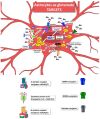
The mechanisms that characterize ALS astrocytes fostering neuronal and glial damage are schematically represented. Neuroinflammation, mitochondrial dysfunction, oxidative stress, energy metabolism impairment, miRNAs and extracellular vesicle involvement, protein misfolding, autophagy dysfunction, and neurotrophic factor dysregulation are major phenomena in ALS progression. The contribution of glutamate and excitotoxicity has been reported in detail in Figures 2 and 3. The figure was generated using BioRender.com (accessed on 25 July 2023) and Servier Medical Art, provided by Servier, licensed under a Creative Commons Attribution 3.0 unported license (https://creativecommons.org/licenses/by/3.0/ (accessed on 25 July 2023)).

Similar articles

Cited by

References

    1. Brown R.H.J. Amyotrophic Lateral Sclerosis: Recent Insights from Genetics and Transgenic Mice. Cell. 1995;80:687–692. doi: 10.1016/0092-8674(95)90346-1. - DOI - PubMed
    1. Brown R.H., Al-Chalabi A. Amyotrophic Lateral Sclerosis. N. Engl. J. Med. 2017;377:162–172. doi: 10.1056/NEJMra1603471. - DOI - PubMed
    1. Dhasmana S., Dhasmana A., Narula A.S., Jaggi M., Yallapu M.M., Chauhan S.C. The Panoramic View of Amyotrophic Lateral Sclerosis: A Fatal Intricate Neurological Disorder. Life Sci. 2022;288:120156. doi: 10.1016/j.lfs.2021.120156. - DOI - PubMed
    1. Phukan J., Pender N.P., Hardiman O. Cognitive Impairment in Amyotrophic Lateral Sclerosis. Lancet Neurol. 2007;6:994–1003. doi: 10.1016/S1474-4422(07)70265-X. - DOI - PubMed
    1. Hardiman O., Al-Chalabi A., Chio A., Corr E.M., Logroscino G., Robberecht W., Shaw P.J., Simmons Z., van den Berg L.H. Amyotrophic Lateral Sclerosis. Nat. Rev. Dis. Primers. 2017;3:17085. doi: 10.1038/nrdp.2017.85. - DOI - PubMed