. 2023 Oct 9;13(10):2031.
doi: 10.3390/life13102031.

Taurine Protects Doxorubicin-Induced Hepatotoxicity via Its Membrane-Stabilizing Effect in Rats

Affiliations

Taurine Protects Doxorubicin-Induced Hepatotoxicity via Its Membrane-Stabilizing Effect in Rats

Esra Gedikli et al. Life (Basel). .

Abstract

Background: Doxorubicin (dox) is a chemotherapeutic agent widely used against various tumors. However, the clinical use of this agent is limited due to various organ toxicities. Taurine is an intracellular free β-amino acid with antioxidant properties. The present study investigated the protective mechanism of taurine on dox-induced hepatotoxicity.

Methods: In total, 31 male Sprague-Dawley rats were used in the study. The control group received intraperitoneal (i.p.) 0.9% NaCl alone for 14 days; the taurine (Tau) group received i.p. taurine 150 mg/kg body weight/day for 14 days; the dox group received dox on days 12, 13, and 14 at a cumulative dose of 25 mg/kg body weight/3 days; and the tau+dox group received taurine and dox together at the same dose and through the same route. On day 15, biochemical evaluations were performed on blood samples taken from the left ventricle followed by histological examinations on liver samples.

Results: Dox was found to increase liver function enzymes and tissue protein carbonyl levels, causing congestion and tissue damage, thereby leading to dysfunction. Tau was found to histologically preserve the liver morphology without showing any corrective effect on oxidative stress parameters. These findings suggest that the membrane-stabilizing effect of taurine may be more effective than its radical scavenging activity in preventing dox-induced toxicity.

Conclusion: Taurine can prevent doxorubicin-induced hepatotoxicity through non-antioxidant pathways.

Keywords: doxorubicin; hepatotoxicity; taurine.


Conflict of interest statement

The authors declare no conflict of interest. The authors have no relevant financial or non-financial interests to disclose. All of the authors declare that they have participated in the design, execution, and analysis of the manuscript and that they have approved the final version.

Figures
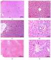
Figure 1
Figure 1
Light microscopic image of liver tissue [H&E]. For all groups; asterisk: central vein. (A) Control group, arrow: hepatocyte cords [×10]. (B) Taurine group, arrow: sinusoidal areas [×20]. (C) Doxorubicin group, arrow: infiltrative cells [×20]. (D) Doxorubicin group, arrow: vacuoles [×40]. (E); Doxorubicin+taurine group [×5], (F). Doxorubicin+taurine group arrow: limiting plate cells [×40].
Figure 2
Figure 2
Electron microscope image of liver tissue. (A) Control group, arrowhead: hepatocyte nucleus, asterisk: Ito cell with fat droplets, and arrow: collagen fibers [×6000]. (B) Taurine group, arrow: hepatocyte nucleus and asterisk: Kupffer cell nucleus [×2500]. (C) Doxorubicin group, rER: tough endoplasmic reticulum and arrow: rER [×8000]. (D) Doxorubicin group, arrow: nuclear pore and asterisk: fat vacuoles [×12,000]. (E) Doxorubicin+taurine group, arrow: rER [×8000]. (F) Doxorubicin+taurine group, arrow: rER and asterisk: heterophagic vacuoles. [×15,000]. rER: rough endoplasmic reticulum.
Figure 2
Figure 2
Electron microscope image of liver tissue. (A) Control group, arrowhead: hepatocyte nucleus, asterisk: Ito cell with fat droplets, and arrow: collagen fibers [×6000]. (B) Taurine group, arrow: hepatocyte nucleus and asterisk: Kupffer cell nucleus [×2500]. (C) Doxorubicin group, rER: tough endoplasmic reticulum and arrow: rER [×8000]. (D) Doxorubicin group, arrow: nuclear pore and asterisk: fat vacuoles [×12,000]. (E) Doxorubicin+taurine group, arrow: rER [×8000]. (F) Doxorubicin+taurine group, arrow: rER and asterisk: heterophagic vacuoles. [×15,000]. rER: rough endoplasmic reticulum.

Similar articles

Cited by

References

    1. Prasanna P.L., Renu K., Gopalakrishnan A.V. New molecular and biochemical insights of doxorubicin-induced hepatotoxicity. Life Sci. 2020;250:117599. doi: 10.1016/j.lfs.2020.117599. - DOI - PubMed
    1. Shokrzadeh M., Bagheri A., Ghassemi-Barghi N., Rahmanian N., Eskandani M. Doxorubicin and doxorubicin-loaded nanoliposome induce senescence by enhancing oxidative stress, hepatotoxicity, and in vivo genotoxicity in male Wistar rats. Naunyn-Schmiedeberg’s Arch. Pharmacol. 2021;394:1803–1813. doi: 10.1007/s00210-021-02119-w. - DOI - PubMed
    1. Liu X., Qiu Y., Liu Y., Huang N., Hua C., Wang Q., Wu Z., Lu J., Song P., Xu J., et al. Citronellal ameliorates doxorubicin-induced hepatotoxicity via antioxidative stress, antiapoptosis, and proangiogenesis in rats. J. Biochem. Mol. Toxicol. 2020;35:e22639. doi: 10.1002/jbt.22639. - DOI - PubMed
    1. Karaman A., Fadillioglu E., Turkmen E., Tas E., Yilmaz Z. Protective effects of leflunomide against ischemia-reperfusion injury of the rat liver. Pediatr. Surg. Int. 2006;22:428–434. doi: 10.1007/s00383-006-1668-x. - DOI - PubMed
    1. Marchand D.J., Renton K.W. Depression of cytochrome P-450-dependent drug biotransformation by adriamycin. Toxicol. Appl. Pharmacol. 1981;58:83–88. doi: 10.1016/0041-008X(81)90118-6. - DOI - PubMed

LinkOut - more resources