

Multiple Primary Melanoma: A Five-Year Prospective Single-Center Follow-Up Study of Two MC1R R/R Genotype Carriers
- PMID: 37895483
- PMCID: PMC10608495
- DOI: 10.3390/life13102102
Multiple Primary Melanoma: A Five-Year Prospective Single-Center Follow-Up Study of Two MC1R R/R Genotype Carriers
Abstract
Background: Multiple primary melanoma (MPM) is a diagnostic challenge even with ancillary imaging technologies available to dermatologists. In selected patients' phenotypes, the use of imaging approaches can help better understand lesion characteristics, and aid in early diagnosis and management.
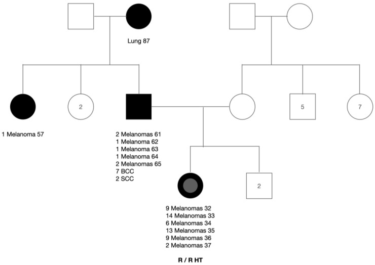
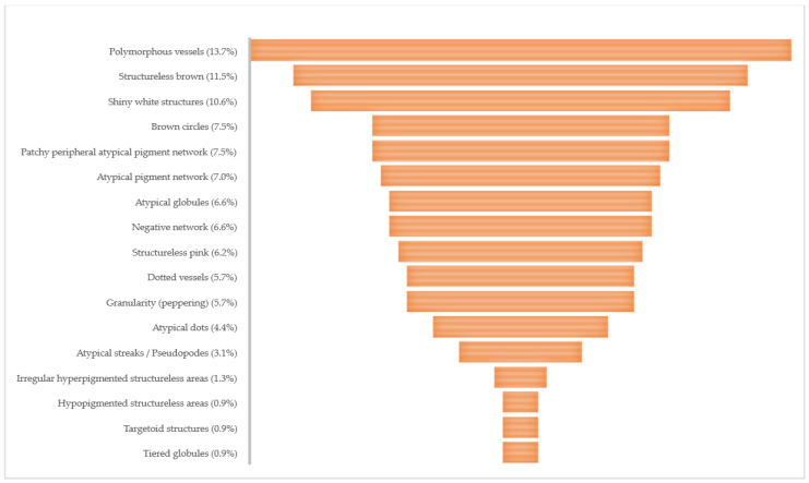
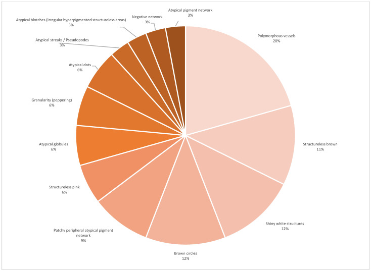
Methods: Under a 5-year prospective single-center follow-up, 58 s primary melanomas (SPMs) were diagnosed in two first-degree relatives, with fair skin color, red hair, green eyes, and personal history of one previous melanoma each. Patients' behavior and descriptive demographic data were collected from medical records. The information on the first two primary melanomas (PMs) were retrieved from pathology reports. The characteristics of 60 melanomas were collected from medical records, video dermoscopy software, and pathology reports. Reflectance confocal microscopy (RCM) was performed prior to excision of 22 randomly selected melanomas.
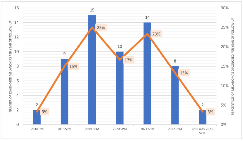
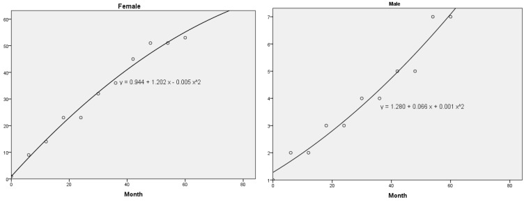
Results: From February 2018 to May 2023, two patients underwent a pooled total of 214 excisional biopsies of suspect lesions, resulting in a combined benign versus malignant treatment ratio (NNT) of 2.0:1.0. The number of moles excised for each melanoma diagnosed (NNE) was 1.7:1.0 and 6.9:1.0 for the female and male patient respectively. The in-situ melanoma/invasive melanoma ratio (IIR) demonstrated a higher proportion of in-situ melanomas for both patients. From June 2018 to May 2023, a total of 58 SPMs were detected by the combination of total body skin exam (TBSE), total body skin photography (TBSP), digital dermoscopy (DD), and sequential digital dermoscopy imaging (SDDI) via comparative approach. The younger patient had her PM one month prior to the second and third cutaneous melanomas (CMs), characterizing a case of synchronous primary CM. The male older relative had a total of 7 nonsynchronous melanomas.
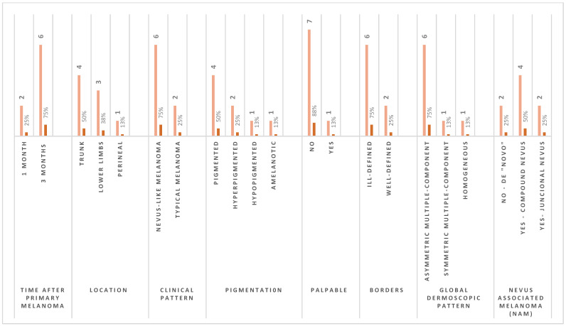
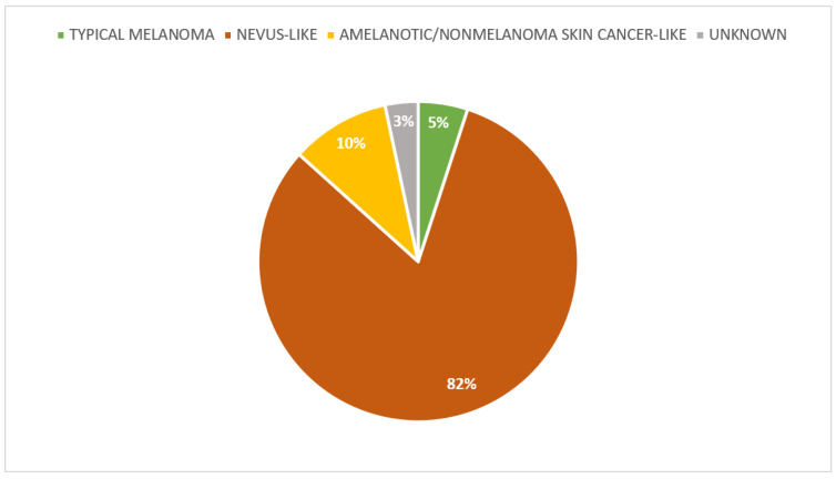
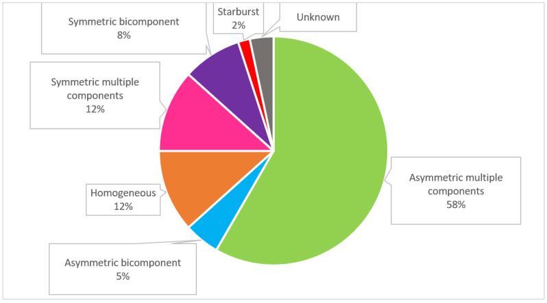
Conclusions: This CM cohort is composed of 83.3% in-situ melanoma and 16.7% invasive melanoma. Both patients had a higher percentage of SPM with clinical nevus-like morphology (84.5%), global dermoscopic pattern of asymmetric multiple component (60.3%) and located on the lower limbs (46.6%). When RCM was performed prior to excision, 81% of SPM had features suggestive of malignancy. As well, invasive melanomas were more frequent in the lower limbs (40%). In the multivariate model, for the two high-risk patients studied, the chance of a not associated with nevus ("de novo") invasive SPM diagnosis is 25 times greater than the chance of a diagnosis of a nevus-associated invasive SPM.
Keywords: MC1R; digital dermoscopy; dysplastic nevus syndrome; melanoma; multiple primary melanoma; nevus associated melanoma; reflectance confocal microscopy; second primary melanoma; sequential digital dermoscopy imaging; skin cancer; synchronous primary cutaneous melanomas; total body skin photograph.
Conflict of interest statement
The authors declare no conflict of interest.
Figures

Similar articles
-
Role of In Vivo Reflectance Confocal Microscopy in the Analysis of Melanocytic Lesions.Acta Dermatovenerol Croat. 2018 Apr;26(1):64-67. Acta Dermatovenerol Croat. 2018. PMID: 29782304 Review.
-
Detection of primary melanoma in individuals at extreme high risk: a prospective 5-year follow-up study.JAMA Dermatol. 2014 Aug;150(8):819-27. doi: 10.1001/jamadermatol.2014.514. JAMA Dermatol. 2014. PMID: 24964862
-
Three-Dimensional Total Body Photography, Digital Dermoscopy, and in vivo Reflectance Confocal Microscopy for Follow-Up Assessments of High-Risk Patients for Melanoma: A Prospective, Controlled Study.Dermatology. 2024;240(5-6):803-813. doi: 10.1159/000541894. Epub 2024 Oct 8. Dermatology. 2024. PMID: 39378859 Free PMC article.
-
Combined in vivo reflectance confocal microscopy and digital dermoscopy for follow up of patients at high risk of malignant melanoma: A prospective case series study.J Dermatol. 2017 Jun;44(6):681-689. doi: 10.1111/1346-8138.13743. Epub 2017 Feb 13. J Dermatol. 2017. PMID: 28191661
-
Surveillance of patients at high risk for cutaneous malignant melanoma using digital dermoscopy.Br J Dermatol. 2005 Jan;152(1):87-92. doi: 10.1111/j.1365-2133.2005.06370.x. Br J Dermatol. 2005. PMID: 15656806 Review.
References
-
- Antúnez-Lay A., Podlipnik S., Carrera C., Potrony M., Tell-Martí G., Badenas C., Puig-Butille J.A., Espinosa N., Puig S., Malvehy J. Synchronous primary cutaneous melanomas: A descriptive study of their clinical features, histology, genetic background of the patients and clinical outcomes. J. Eur. Acad. Dermatol. Venereol. 2022;36:2364–2372. doi: 10.1111/jdv.18467. - DOI - PubMed
-
- Russo T., Piccolo V., Moscarella E., Tschandl P., Kittler H., Paoli J., Lallas A., Braun R.P., Thomas L., Soyer H.P., et al. Indications for Digital Monitoring of Patients with Multiple Nevi: Recommendations from the International Dermoscopy Society. Dermatol. Pract. Concept. 2022;12:e2022182. doi: 10.5826/dpc.1204a182. - DOI - PMC - PubMed
-
- Elder D.E., Massi D., Scolyer R.A., Willemze R. WHO Classification of Skin Tumours. 4th ed. WHO; Geneva, Switzerland: 2018.
LinkOut - more resources
Full Text Sources
Research Materials
