. 2023 Sep 28;16(10):1379.
doi: 10.3390/ph16101379.

Unlocking the Power of Onion Peel Extracts: Antimicrobial and Anti-Inflammatory Effects Improve Wound Healing through Repressing Notch-1/NLRP3/Caspase-1 Signaling

Affiliations

Unlocking the Power of Onion Peel Extracts: Antimicrobial and Anti-Inflammatory Effects Improve Wound Healing through Repressing Notch-1/NLRP3/Caspase-1 Signaling

Rafik Mounir et al. Pharmaceuticals (Basel). .

Abstract

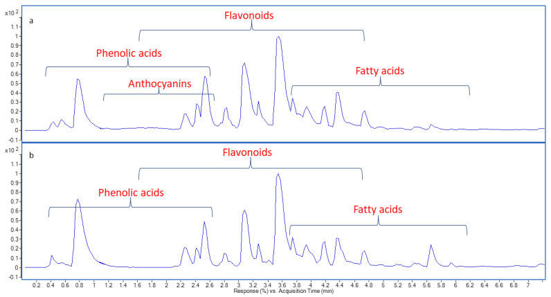
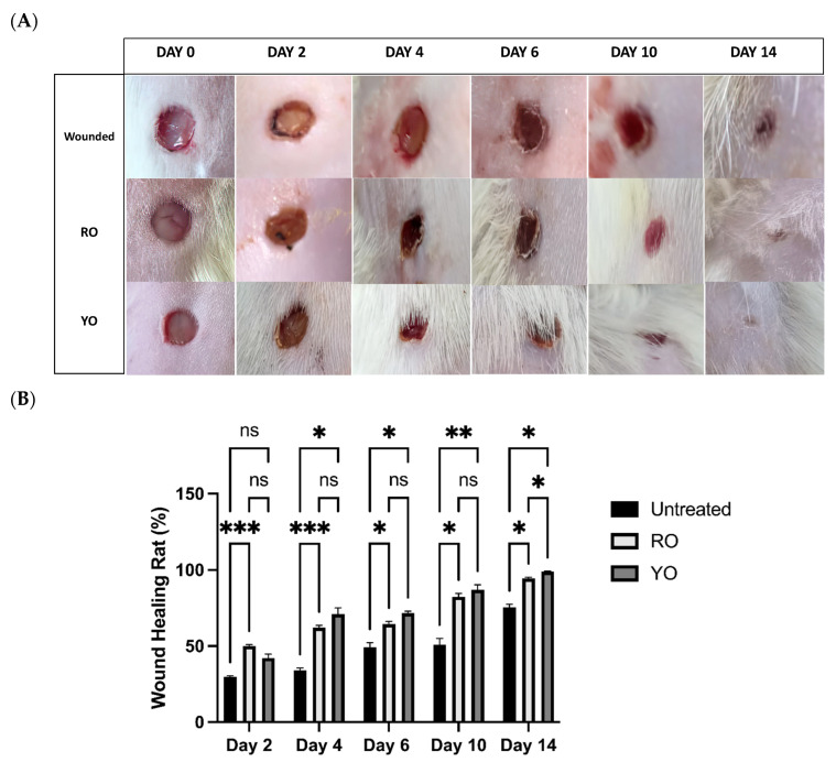
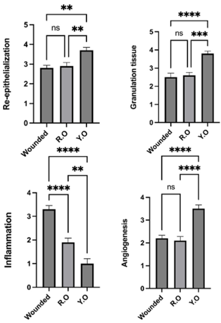
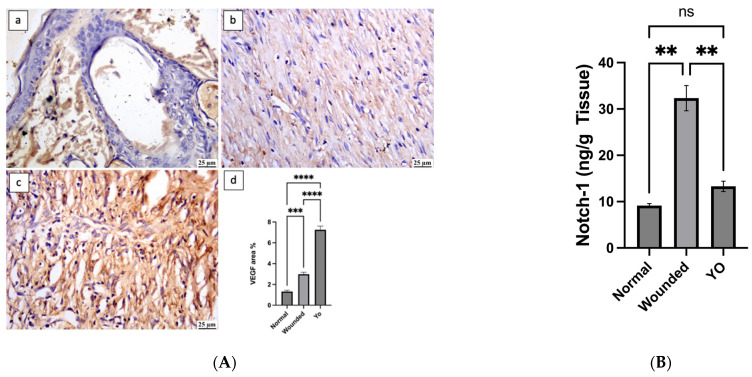
Onion peels are often discarded, representing an unlimited amount of food by-products; however, they are a valuable source of bioactive phenolics. Thus, we utilized UPLC-MS/MS to analyze the metabolomic profiles of red (RO) and yellow (YO) onion peel extracts. The cytotoxic (SRB assay), anti-inflammatory (Griess assay), and antimicrobial (sensitivity test, MIC, antibiofilm, and SP-SDS tests) properties were assessed in vitro. Additionally, histological analysis, immunohistochemistry, and ELISA tests were conducted to investigate the healing potential in excisional skin wound injury and Candida albicans infection in vivo. RO extract demonstrated antibacterial activity, limited skin infection with C. albicans, and improved the skin's appearance due to the abundance of quercetin and anthocyanin derivatives. Both extracts reduced lipopolysaccharide-induced nitric oxide release in vitro and showed a negligible cytotoxic effect on MCF-7 and HT29 cells. When extracts were tested in vivo for their ability to promote tissue regeneration, it was found that YO peel extract had the greatest impact. Further biochemical analysis revealed that YO extract suppressed NLRP3/caspase-1 signaling and decreased inflammatory cytokines. Furthermore, YO extract decreased Notch-1 levels and boosted VEGF-mediated angiogenesis. Our findings imply that onion peel extract can effectively treat wounds by reducing microbial infection, reducing inflammation, and promoting tissue regeneration.

Keywords: Candida; MRSA; VEGF; angiogenesis; anthocyanins; dietary flavonoids; quercetin; tissue regeneration.


Conflict of interest statement

The authors declare no conflict of interest.

Figures

Figure 1
Figure 1
Base peak chromatograms of onion peels; RO (a) and YO (b) methanol extracts in negative ionization mode.
Figure 2
Figure 2
Mass fragments of some identified phytoconstituents in the onion peel extract: (a) quercetin, O-(O-malonyl-hexoside), O-hexoside; (b) quercetin-O-acetylhexoside.
Figure 3
Figure 3
Demonstration of biofilm reduction and destructive effects of RO peel extract on microbial isolates at concentrations of MIC, 2MIC, and 4MIC.
Figure 4
Figure 4
(A): In vivo imaging of rat skin infected with Candida albicans. (B): The size of infected skin by C. albicans when analyzed by ImageJ software. Significance is considered at p < 0.05. The quantity of asterisks (*) attached to the columns denotes the degree of significance; ns: no significance. Data are presented as mean ± SEM.
Figure 5
Figure 5
Wound healing and tissue repair in rats treated with RO and YO peel extracts. (A): Optical images; (B): Wound healing rate % at 0, 2, 4, 6, 10, and 14 days. Data are presented as mean ± SEM. Significance is considered at p < 0.05. The quantity of asterisks (*) denotes the degree of significance; (*): p < 0.05, (**): p < 0.01, (***): p < 0.001; ns: no significance.
Figure 6
Figure 6
Photomicrographs of skin (H&E) showing (a) negative control group: normal skin; (b) wounded group: wound area filled with granulation tissue and complete re-epithelization; (c) RO-treated group: filling of wound area with collagen-rich tissue and complete re-epithelization; (d) YO-treated group: contracture of the wound area with complete re-epithelization.
Figure 7
Figure 7
Wound healing histological score: charts represent healing criteria in each group. The negative control group showed normal skin without wound area. Data are presented as mean ± SEM. Significance is considered at p < 0.05. The quantity of asterisks (*) denotes the degree of significance; (**): p < 0.01, (***): p < 0.001, (****): p < 0.0001; ns: no significance.
Figure 8
Figure 8
Photomicrograph of skin, immune expression of NF-κβ (immune staining). (a) Normal group showing limited NF-κβ expression; (b) wounded group showing intense NF-κβ expression; (c) YO-treated group showing moderate NF-κβ expression; (d) chart representing NF-κβ expression. Data are presented as mean ± SEM. Significance is considered at p < 0.05. The quantity of asterisks (*) denotes the degree of significance; (****): p < 0.0001; ns: no significance.
Figure 9
Figure 9
Photomicrograph of skin, immune expression of TNF-α (immune staining). (a) Normal group showing weak TNF-α expression; (b) wounded group showing increased TNF-α expression; (c) YO-treated group showing moderate TNF-α expression; (d) chart representing TNF-α expression. Data are presented as mean ± SEM. Significance is considered at p < 0.05. The quantity of asterisks (*) denotes the degree of significance; (****): p < 0.0001; ns: no significance.
Figure 10
Figure 10
Photomicrograph of skin, immune expression of IL-1β (immune staining). (a) Normal group showing weak IL-1β expression; (b) wounded group showing intense IL-1β expression; (c) YO-treated group showing moderate IL-1β expression; (d) chart representing IL-1β expression. Data are presented as mean ± SEM. Significance is considered at p < 0.05. The quantity of asterisks (*) denotes the degree of significance; (****): p < 0.0001; ns: no significance.
Figure 11
Figure 11
(A): Photomicrograph of skin, immune expression of VEGF (immune staining). (a) Normal group showing mild VEGF expression; (b) wounded group showing moderate VEGF expression; (c) YO-treated group showing intense VEGF expression; (d) chart representing VEGF expression. (B): Notch-1 levels (ng/g tissue) in normal, wounded, and YO-treated groups. Data are presented as mean ± SEM. Significance is considered at p < 0.05. The quantity of asterisks (*) denotes the degree of significance; (**): p < 0.01, (***): p < 0.001, (****): p < 0.0001; ns: no significance.
Figure 12
Figure 12
(A): Caspase-1 level (ng/g tissue) in normal, wounded, and YO-treated groups. (B): NLRP3 level (ng/g tissue) in normal, wounded, and YO-treated groups. Data are presented as mean ± SEM. Significance is considered at p < 0.05. The quantity of asterisks (*) denotes the degree of significance; (**): p < 0.01, (***): p < 0.001, (****): p < 0.0001; ns: no significance.

References

    1. Suleria H.A.R., Butt M.S., Anjum F.M., Saeed F., Khalid N. Onion: Nature protection against physiological threats. Crit. Rev. Food Sci. Nutr. 2015;55:50–66. doi: 10.1080/10408398.2011.646364. - DOI - PubMed
    1. Griffiths G., Trueman L., Crowther T., Thomas B., Smith B. Onions—A global benefit to health. Phyther. Res. 2002;16:603–615. doi: 10.1002/ptr.1222. - DOI - PubMed
    1. FAOSTAT FAO Statistics Division. 2018. [(accessed on 1 May 2022)]. Available online: https://www.fao.org/faostat/en/#data.
    1. Omar A.E., Al-Khalaifah H.S., Mohamed W.A.M., Gharib H.S.A., Osman A., Al-Gabri N.A., Amer S.A. Effects of Phenolic-Rich Onion (Allium cepa L.) Extract on the Growth Performance, Behavior, Intestinal Histology, Amino Acid Digestibility, Antioxidant Activity, and the Immune Status of Broiler Chickens. Front. Vet. Sci. 2020;7:582612. doi: 10.3389/fvets.2020.582612. - DOI - PMC - PubMed
    1. Colina-Coca C., González-Peña D., de Ancos B., Sánchez-Moreno C. Dietary onion ameliorates antioxidant defence, inflammatory response, and cardiovascular risk biomarkers in hypercholesterolemic Wistar rats. J. Funct. Foods. 2017;36:300–309. doi: 10.1016/j.jff.2017.07.014. - DOI

LinkOut - more resources