. 2023 Oct 12;16(10):1446.
doi: 10.3390/ph16101446.

Spironolactone Eyedrop Favors Restoration of Corneal Integrity after Wound Healing in the Rat

Affiliations

Spironolactone Eyedrop Favors Restoration of Corneal Integrity after Wound Healing in the Rat

Daniela Rodrigues-Braz et al. Pharmaceuticals (Basel). .

Abstract

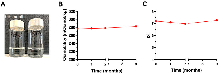
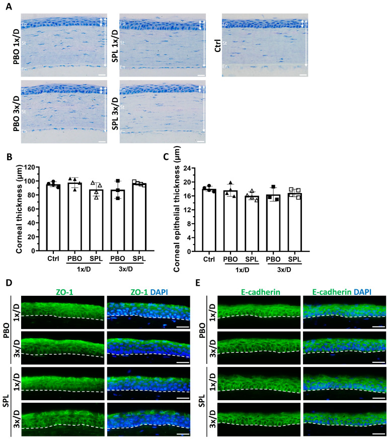
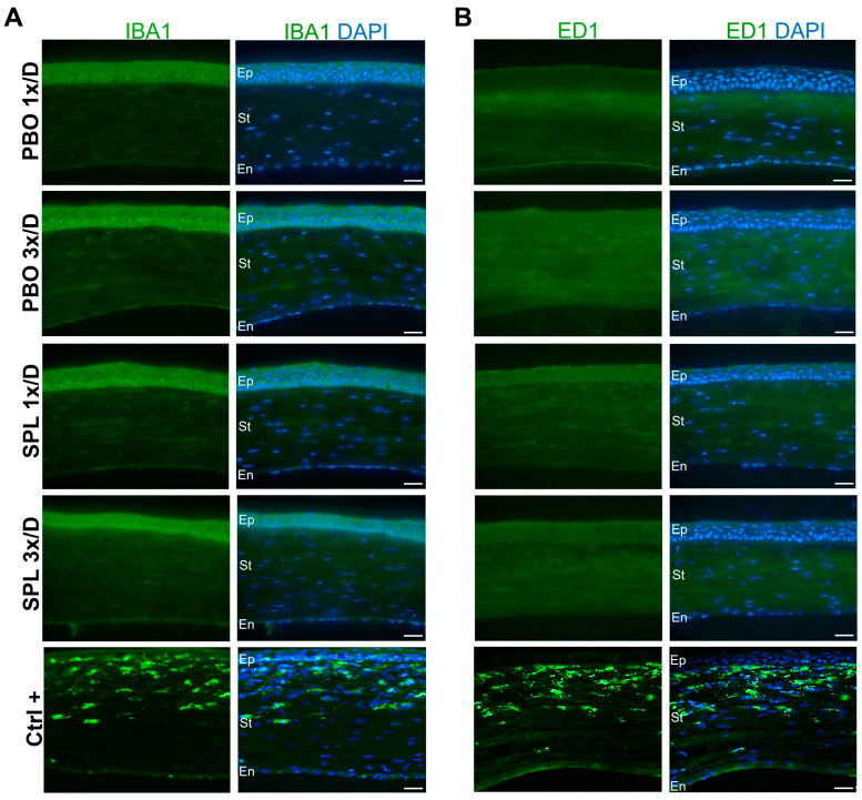
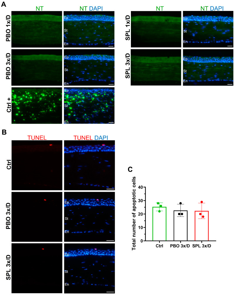
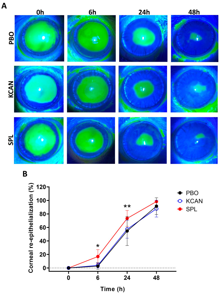
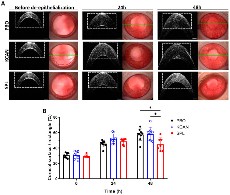
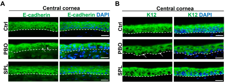
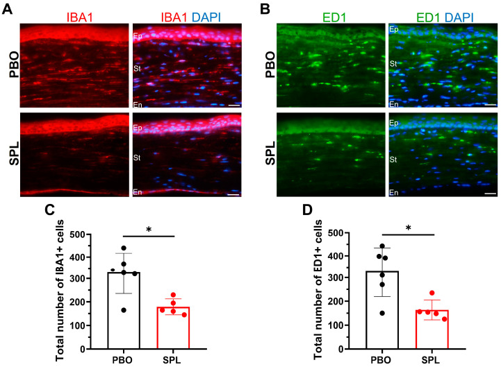
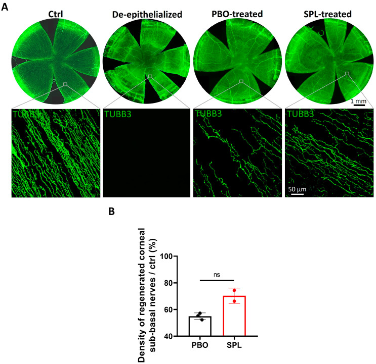
Abnormal corneal wound healing can compromise corneal transparency and lead to visual impairment. Mineralocorticoid receptor antagonists (MRA) are promising candidates to promote corneal remodeling with anti-inflammatory properties and lack gluococorticoids-associated side effects. In this preclinical study, a new polymer-free hydroxypropyl-gamma-cyclodextrin-based eyedrop containing 0.1% spironolactone (SPL), a potent but non-water-soluble MRA, was investigated for its ocular surface tolerance and efficacy in a rat model of corneal wound healing. SPL eyedrops were stable for up to 9 months at 4 °C. The formulation was well-tolerated since no morphological changes or inflammatory reactions were observed in the rat cornea after multiple daily instillations over 7 days. SPL eyedrops accelerated rat corneal wound healing, reduced corneal edema and inflammation, enhanced epithelial integrity, and improved nerve regeneration, suggesting restoration of corneal homeostasis, while potassium canrenoate, an active and soluble metabolite of SPL, had no effect. SPL eyedrops could benefit patients with impaired corneal wound healing, including that secondary to glucocorticoid therapy. Repurposing known drugs with known excipients will expedite translation to the clinic.

Keywords: cornea; corneal wound healing; drug development; eyedrop; ocular surface; spironolactone; topical administration.


Conflict of interest statement

J.-P.C. and X.C. are employees of Excelvision, Fareva, Annonay, France. Y.B. is an employee of Valdepharm, Fareva, Val-de-Reuil, France. P.R. is an employee of Fareva, France. D.R.-B., L.Z., E.G., A.T., J.-L.B., F.B.-C. and M.Z. declare no conflict of interest.

Figures

Figure 1
Figure 1
Stability of eyedrop formulations. (A) Spironolactone (SPL) and placebo (PBO) eyedrops remain clear and colorless after 9 months’ storage at 4 °C. (B) The osmolality of SPL eyedrops over time up to 9 months. (C) The pH of SPL eyedrops over time up to 9 months.
Figure 2
Figure 2
Rat corneal and corneal epithelial morphology after topical instillation of eyedrops containing spironolactone for 7 days. (A) Histological sections show normal corneal structure with stratified corneal epithelium (Ep), well-organized stroma (St), and intact corneal endothelial (En) monolayer after instillation of eyedrops containing 0.1% spironolactone (SPL) or placebo (PBO) once or three times a day for 7 days, similar to that of the untreated control cornea (Ctrl). Double-headed white arrows indicate the thickness of the cornea and corneal epithelium. Scale bars: 20 µm. (B) Quantification of the corneal thickness on histological sections shows no significant difference among all groups. Data are expressed as mean ± SD, n = 3–4 rat corneas. (C) Quantification of the corneal epithelial thickness shows no significant difference among all groups. Data are expressed as mean ± SD, n = 3–4 rat corneas. (D) ZO-1 immunofluorescence in green shows a cell membrane localization mainly in the superficial layers of the corneal epithelium. No disruption of the immunostaining was observed after topical treatment with SPL and PBO eyedrops. Dash lines indicate the inner limit of the corneal epithelium. Nuclei were counter-stained with DAPI in blue. Scale bars: 20 µm. n = 3–4 rat corneas. (E) E-cadherin immunofluorescence in green shows a cell membrane localization in all epithelial cells. No disruption of the immunostaining was observed after topical treatment with SPL or PBO eyedrops. Dash lines indicate the inner limit of the corneal epithelium. Nuclei were counter-stained with DAPI in blue. Scale bars: 20 µm. n = 3–4 rat corneas.
Figure 3
Figure 3
Immunostaining of inflammatory cells in rat corneas after topical instillation of eyedrops containing spironolactone for 7 days. (A,B) No IBA1- or ED1-positive inflammatory cells were observed in corneas treated with eyedrops containing 0.1% spironolactone (SPL) or placebo (PBO) once or 3 times a day for 7 days. Positive control corneas (Ctrl+) from an inflammation model show IBA and ED1 staining in green in inflamed corneas. Nuclei were counter-stained with DAPI in blue. Scale. Ep, epithelium; St, stroma; En, endothelium. Scale bars: 20 µm. n = 3–4 rat corneas.
Figure 4
Figure 4
Nitrosative stress and cell death markers in rat corneas after topical instillation of eyedrops containing spironolactone for 7 days. (A) No nitrotyrosine (NT) positive cells were observed in corneas treated with eyedrops containing 0.1% spironolactone (SPL) or placebo (PBO) once or 3 times a day for 7 days. Positive control corneas (Ctrl+) from an inflammation model show NT staining in green. Nuclei were counter-stained with DAPI in blue. Ep, epithelium; St, stroma; En, endothelium. Scale bars: 20 µm. n = 3–4 rat corneas. (B) TUNEL positive apoptotic cells in red are only present in the superficial layers of the corneal epithelium in untreated control eyes (Ctrl) as well as in eyes treated with SPL or PBO eyedrops 3 times a day for 7 days. Nuclei were counter-stained with DAPI in blue. Scale bars: 20 µm. (C) Quantification of TUNEL positive cells across the whole corneal epithelium shows no significant differences amongst Ctrl, PBO, and SPL groups. Data are expressed as mean ± SD, n = 3 rat corneas.
Figure 5
Figure 5
SPL eyedrops improve rat corneal re-epithelialization. (A) Fluorescein staining shows the 4-mm central corneal epithelial wound created at 0 h, and healing progress at 6, 24, and 48 h. Spironolactone (SPL) eyedrops improve corneal re-epithelialization compared to the placebo (PBO) and potassium canrenoate solution (KCAN). (B) Quantitative analysis shows that SPL eyedrops significantly increase the epithelial healing rate at 6 and 24 h compared with the PBO eyedrops and KCAN solution, while there is no difference between the KCAN and PBO groups. Data are expressed as mean ± SD, n = 7–9 rat corneas. *, p < 0.05; **, p < 0.01.
Figure 6
Figure 6
SPL eyedrops reduce rat corneal edema after corneal de-epithelialization. (A) In vivo B-scan optical coherence tomography shows the cross-sectional images (left) of the central cornea through the green line (right) before and 24 and 48 h after corneal de-epithelialization. Spironolactone (SPL) eyedrops reduce the corneal edema at 48 h as compared to placebo eyedrops (PBO) and potassium canrenoate solution (KCAN). The rectangles are of the same size and were used as a reference for quantitative analysis of corneal edema. (B) Corneal edema is presented as a percentage of the area of the reference rectangle occupied by the cornea. SPL eyedrops decrease corneal edema significantly compared to the PBO and KCAN groups at 48 h. Data are expressed as mean ± SD, n = 7–9 rat corneas. *, p < 0.05.
Figure 7
Figure 7
SPL eyedrops restore corneal epithelial integrity in rats after corneal wound healing. (A) Immunostaining of E-cadherin (green) shows discontinuity (arrows) and disorganization of E-cadherin in the epithelium of placebo-treated (PBO) corneas. Spironolactone eyedrops (SPL) restore E-cadherin continuity and the stratified structure of the corneal epithelium comparable to the control uninjured cornea (Ctrl). Dash lines indicate the inner border of the corneal epithelium. Nuclei were counter-stained with DAPI in blue. Scale bars: 20 µm. n = 4–5 rat corneas. (B) Immunostaining of cytokeratin 12 (K12, green) shows a decrease in K12 fluorescence and disruption (arrows) in the basal layer of the epithelium of PBO-treated corneas. The inner edge of the epithelium (dash line) is irregular. SPL eyedrops enhance K12 expression in the corneal epithelium and improve the regularity of the epithelial inner border, comparable to the control cornea. Nuclei were counter-stained with DAPI in blue. Scale bars: 20 µm. n = 4–5 rat corneas.
Figure 8
Figure 8
SPL eyedrops reduce inflammatory cell infiltration in rat corneas after corneal de-epithelialization. (A,B) IBA1 (in red) and ED1 immunostaining (in green) shows infiltration of inflammatory cells throughout the stroma of placebo-treated (PBO) corneas, whereas spironolactone eyedrops (SPL) limit the cell infiltration in the anterior stroma. Nuclei were counter-stained with DAPI in blue. Dash lines indicate the inner border of the corneal epithelium. Ep, epithelium; St, stroma; En, endothelium. Scale bars: 20 µm. (C,D) SPL eyedrops significantly decrease the number of IBA1- and ED1-positive cells in rat corneas compared with the PBO-treated group. Data are expressed as mean ± SD, n = 5–6 rat corneas. *, p < 0.05.
Figure 9
Figure 9
SPL eyedrops favor corneal re-innervation in the de-epithelialized central cornea in rats. (A) TUBB3 immunostaining in green on corneal whole mounts shows dense and linear corneal sub-basal nerves in control uninjured corneas (Ctrl). Corneal de-epithelialization mechanically removed the sub-basal nerve plexus. Spironolactone (SPL) eyedrops improve the morphology of regenerated sub-basal nerves compared with placebo-treated (PBO) corneas. (B) The density of regenerated sub-basal nerves is 54.95% relative to control cornea in the PBO-treated group (n = 3) and increased to 70.32% in the SPL-treated group (n = 2). Data are expressed as mean ± SD; ns, not significant.

Similar articles

Cited by

References

    1. DelMonte D.W., Kim T. Anatomy and Physiology of the Cornea. J. Cataract Refract. Surg. 2011;37:588–598. doi: 10.1016/j.jcrs.2010.12.037. - DOI - PubMed
    1. Labetoulle M., Baudouin C., Calonge M., Merayo-Lloves J., Boboridis K.G., Akova Y.A., Aragona P., Geerling G., Messmer E.M., Benítez-del-Castillo J. Role of Corneal Nerves in Ocular Surface Homeostasis and Disease. Acta Ophthalmol. 2019;97:137–145. doi: 10.1111/aos.13844. - DOI - PubMed
    1. Mohan R.R., Kempuraj D., D’Souza S., Ghosh A. Corneal Stromal Repair and Regeneration. Prog. Retin. Eye Res. 2022;91:101090. doi: 10.1016/j.preteyeres.2022.101090. - DOI - PMC - PubMed
    1. Jeang L., Tuli S.S. Therapy for Contact Lens-Related Ulcers. Curr. Opin. Ophthalmol. 2022;33:282–289. doi: 10.1097/ICU.0000000000000861. - DOI - PubMed
    1. Netto M.V., Mohan R.R., Ambrósio R., Hutcheon A.E.K., Zieske J.D., Wilson S.E. Wound Healing in the Cornea: A Review of Refractive Surgery Complications and New Prospects for Therapy. Cornea. 2005;24:509–522. doi: 10.1097/01.ico.0000151544.23360.17. - DOI - PubMed

LinkOut - more resources