. 2023 Oct 16;16(10):1472.
doi: 10.3390/ph16101472.

Tumor-Infiltrating iNKT Cells Activated through c-Kit/Sca-1 Are Induced by Pentoxifylline, Norcantharidin, and Their Mixtures for Killing Murine Melanoma Cells

Affiliations

Tumor-Infiltrating iNKT Cells Activated through c-Kit/Sca-1 Are Induced by Pentoxifylline, Norcantharidin, and Their Mixtures for Killing Murine Melanoma Cells

Maximiliano V M Correa-Lara et al. Pharmaceuticals (Basel). .

Abstract

The involvement of NK and other cytotoxic cells is considered the first defense line against cancer. However, a significant lack of information prevails on the possible roles played by factors considered characteristic of primitive cells, such as c-kit and Sca-1, in activating these cells, particularly in melanoma models subjected to treatments with substances under investigation, such as the case of norcantharidin. In this study, B16F1 murine melanoma cells were used to induce tumors in DBA/2 mice, estimating the proportions of NK and iNKT cells; the presence of activation (CD107a+) and primitive/activation (c-kit+/Lya6A+) markers and some tumor parameters, such as the presence of mitotic bodies, nuclear factor area, NK and iNKT cell infiltration in the tumor, infiltrated tumor area, and infiltrating lymphocyte count at 10x and 40x in specimens treated with pentoxifylline, norcantharidin, and the combination of both drugs. Possible correlations were estimated with Pearson's correlation analysis. It should be noted that, despite having demonstrated multiple correlations, immaturity/activation markers were related to these cells' activation. At the tumor site, iNKT cells are the ones that exert the cytotoxic potential on tumor cells, but they are confined to specific sites in the tumor. Due to the higher number of interactions of natural killer cells with tumor cells, it is concluded that the most effective treatment was PTX at 60 mg/kg + NCTD at 0.75 mg/kg.

Keywords: CD107a+; Lya6A+; NK; c-kit+; iNKT.


Conflict of interest statement

The authors declare that they have no known competing financial interest or personal relationships that could have appeared to influence the work reported in this manuscript.

Figures
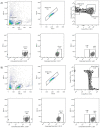
Figure 1
Figure 1
Gating strategy to identify subpopulations of natural killer cells. Panel (A) for NK cells shows from left to right the number of events recorded, number of lymphocytes, number of NK cells (CD56+ and CD16+ positivity), number of active NK cells (CD107+), number of NK cells with the marker c-kit (CD117+), and number of NK cells with the Sca-1 marker (Ly6A+). Panel (B) for iNKT cells shows, from left to right, the number of events recorded, number of lymphocytes, number of iNKT cells (CD1D+ and TCRVB8.1+ positivity), number of active iNKT cells (CD107+), number of iNKT cells with c-kit marker (CD117+), and number of iNKT cells with Sca-1 marker (Ly6A+).
Figure 2
Figure 2
The phenotype of peripheral NK cells. (A) Proportion of NK cells concerning the number of total lymphocytes. (B) Proportion of NK cells (CD107+) to the number of total lymphocytes. (C) Proportion of NK cells (CD117 + LY6A +). Statistical differences to the control, p ≤ 0.05, p ≤ 0.01, and p ≤ 0.001.
Figure 3
Figure 3
The phenotype of peripheral iNKT cells. (A) Proportion of iNKT cells (CD1D + TCRVB8.1+) concerning the number of total lymphocytes. (B) Proportion of iNKT cells (CD107+) to the number of total lymphocytes. (C) Proportion of iNKT cells (CD117 + LY6A +) about the number of total lymphocytes. Statistical differences concerning the control, p ≤ 0.05, p ≤ 0.01, and p ≤ 0.001.
Figure 4
Figure 4
Histopathological analysis. (A) Infiltration of mononuclear cells/10 fields—40x. The treatment group with PTX 60 mg/kg in combination with NCTD 3 mg/kg (v.p.) showed a more significant infiltration of mononuclear cells concerning the control group, with statistical significance (** p ≤ 0.01). (B) Mitotic figures, the treatment groups with PTX 60 mg/kg in combination with NCTD 3 mg/kg and PTX 60 mg/kg in combination with NCTD 0.75 mg/kg showed a lower presence of mitotic bodies to the control group, with statistical significance (** p ≤ 0.01 and * p ≤ 0.05, respectively). (C) Nuclear factor area, the treatment groups with PTX 60 mg/kg in combination with NCTD 3 mg/kg and PTX 60 mg/kg in combination with NCTD 0.75 mg/kg showed a higher nuclear area factor concerning the group control, with statistical significance (** p ≤ 0.001 and * p ≤ 0.05, respectively). (D) Infiltration of mononuclear cell/ImageJ—10x. The treatment group with PTX 60 mg/kg in combination with NCTD 3 mg/kg (v.p.) showed a more significant infiltration of mononuclear cells concerning the control group with statistical significance (** p ≤ 0.01and * p ≤ 0.05, respectively). (E) % Area mononuclear cell/ImageJ—10x. The treatment group with PTX 60 mg/kg in combination with NCTD 3 mg/kg (v.p.) showed a more significant infiltration of mononuclear cells concerning the control group with statistical significance (** p ≤ 0.01and * p ≤ 0.05, respectively). ns: not significant.
Figure 5
Figure 5
Immunofluorescence microscopy image of merged NK cells (CD16+/CD56+/DAPI+) by treatment group. (A) Immunofluorescence microscopy analysis, of intratumoral lymphocytic infiltration by NK cells (CD16 + CD56+) with statistical significance for the treatment group with PTX 60 mg/kg in combination with NCTD 3 mg/kg (*** p ≤ 0.001). No statistically significant difference was detected in the rest of the treatments. (B) Control group, sterile saline solution. (C) PTX 60 mg/kg. (D) PTX 30 mg/kg. (E) NCTD 3 mg/kg. (F) PTX 60 mg/kg + NCTD 3 mg/kg. (G) PTX 60 mg + NCTD 0.75 mg/kg. (H) NCTD 0.75 mg/kg. ns: not significant.
Figure 6
Figure 6
Immunofluorescence microscopy image of iNKT cells in merged (CD1D+/TCRVB8.1+/DAPI+) by treatment group. (A) Immunofluorescence microscopy analysis of intratumoral lymphocytic infiltration by iNKT cells (CD1D + TCRVB8.1+) with statistical significance for the treatment group with PTX 60 mg/kg in combination with NCTD 3 mg/kg, (* p < 0.05). (B) Control group, sterile saline solution. (C) PTX 60 mg/kg. (D) PTX 30 mg/kg. (E) NCTD 3 mg/kg. (F) PTX 60 mg/kg + NCTD 3 mg/kg. (G) PTX 60 mg + NCTD 0.75 mg/kg. (H) NCTD 0.75 mg/kg. ns: not significant.
Figure 7
Figure 7
The figure shows the days of drug administration: pentoxifylline was administered on days 2, 4, 6, 7, and 8; norcantharidin was administered on days 1, 3, 5, 7, and 8.
Figure 8
Figure 8
Graphic representation of the correlations of the natural killer cell subpopulations (NK and iNKT) in peripheral blood and at the tumor site in a mouse melanoma model treated with pentoxifylline (PTX), norcantharidin (NCTD), and their mixtures. (A) Peripheral blood. According to the evaluation by flow cytometry, the following distribution groups of the subpopulations of NK and iNKT cells were formed. (B) Melanoma tumor infiltration of NK and iNKT cells by Confocal microscope.

Similar articles

Cited by

References

    1. Lanier L.L. Nk cell recognition. Annu. Rev. Immunol. 2005;23:225–274. doi: 10.1146/annurev.immunol.23.021704.115526. - DOI - PubMed
    1. Spits H., Artis D., Colonna M., Diefenbach A., Di Santo J.P., Eberl G., Koyasu S., Locksley R.M., McKenzie A.N.J., Mebius R.E., et al. Innate lymphoid cells—A proposal for uniform nomenclature. Nat. Rev. Immunol. 2013;13:145–149. doi: 10.1038/nri3365. - DOI - PubMed
    1. Guillerey C., Huntington N.D., Smyth M.J. Targeting natural killer cells in cancer immunotherapy. Nat. Immunol. 2016;17:1025–1036. doi: 10.1038/ni.3518. - DOI - PubMed
    1. O’Sullivan T.E. Dazed and confused: NK cells. Front. Immunol. 2019;10:2235. doi: 10.3389/fimmu.2019.02235. - DOI - PMC - PubMed
    1. Cui J., Shin T., Kawano T., Sato H., Kondo E., Toura I., Kaneko Y., Koseki H., Kanno M., Taniguchi M. Requirement for Vα14 NKT cells in IL-12-mediated rejection of tumors. Science. 1997;278:1623–1626. doi: 10.1126/science.278.5343.1623. - DOI - PubMed