1-Piperidine Propionic Acid as an Allosteric Inhibitor of Protease Activated Receptor-2
- PMID: 37895957
- PMCID: PMC10610151
- DOI: 10.3390/ph16101486
1-Piperidine Propionic Acid as an Allosteric Inhibitor of Protease Activated Receptor-2
Abstract
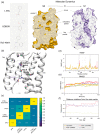
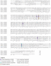
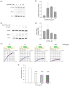
In the last decades, studies on the inflammatory signaling pathways in multiple pathological contexts have revealed new targets for novel therapies. Among the family of G-protein-coupled Proteases Activated Receptors, PAR2 was identified as a driver of the inflammatory cascade in many pathologies, ranging from autoimmune disease to cancer metastasis. For this reason, many efforts have been focused on the development of potential antagonists of PAR2 activity. This work focuses on a small molecule, 1-Piperidine Propionic Acid (1-PPA), previously described to be active against inflammatory processes, but whose target is still unknown. Stabilization effects observed by cellular thermal shift assay coupled to in-silico investigations, including molecular docking and molecular dynamics simulations, suggested that 1-PPA binds PAR2 in an allosteric pocket of the receptor inactive conformation. Functional studies revealed the antagonist effects on MAPKs signaling and on platelet aggregation, processes mediated by PAR family members, including PAR2. Since the allosteric pocket binding 1-PPA is highly conserved in all the members of the PAR family, the evidence reported here suggests that 1-PPA could represent a promising new small molecule targeting PARs with antagonistic activity.
Keywords: 1-piperidinepropionic acid; G protein-coupled receptors; Protease Activated Receptor 2; allosteric modulator; molecular dynamics.
Conflict of interest statement
Pontisso P, Biasiolo A, Cendron L, Chinellato M, Quarta, S, Ruvoletto M. are inventors of the Patent Application of the University of Padova N. 102022000014593. No conflict of interest exists for the other authors.
Figures


Similar articles
-
Structural insight into allosteric modulation of protease-activated receptor 2.Nature. 2017 May 4;545(7652):112-115. doi: 10.1038/nature22309. Epub 2017 Apr 26. Nature. 2017. PMID: 28445455
-
Pharmacophore-based virtual screening, biological evaluation and binding mode analysis of a novel protease-activated receptor 2 antagonist.J Comput Aided Mol Des. 2016 Aug;30(8):625-37. doi: 10.1007/s10822-016-9937-9. Epub 2016 Sep 6. J Comput Aided Mol Des. 2016. PMID: 27600555
-
Protease-activated receptor-2 ligands reveal orthosteric and allosteric mechanisms of receptor inhibition.Commun Biol. 2020 Dec 17;3(1):782. doi: 10.1038/s42003-020-01504-0. Commun Biol. 2020. PMID: 33335291 Free PMC article.
-
Allosteric modulation of protease-activated receptor signaling.Mini Rev Med Chem. 2012 Aug;12(9):804-11. doi: 10.2174/138955712800959116. Mini Rev Med Chem. 2012. PMID: 22681248 Free PMC article. Review.
-
The development of proteinase-activated receptor-2 modulators and the challenges involved.Biochem Soc Trans. 2020 Dec 18;48(6):2525-2537. doi: 10.1042/BST20200191. Biochem Soc Trans. 2020. PMID: 33242065 Free PMC article. Review.
Cited by
-
1-Piperidine Propionic Acid Protects from Septic Shock Through Protease Receptor 2 Inhibition.Int J Mol Sci. 2024 Oct 30;25(21):11662. doi: 10.3390/ijms252111662. Int J Mol Sci. 2024. PMID: 39519216 Free PMC article.
-
Inhibition of Protease-Activated Receptor-2 Activation in Parkinson's Disease Using 1-Piperidin Propionic Acid.Biomedicines. 2024 Jul 22;12(7):1623. doi: 10.3390/biomedicines12071623. Biomedicines. 2024. PMID: 39062196 Free PMC article.
-
Protease activated receptor 2 as a novel druggable target for the treatment of metabolic dysfunction-associated fatty liver disease and cancer.Front Immunol. 2024 Oct 11;15:1397441. doi: 10.3389/fimmu.2024.1397441. eCollection 2024. Front Immunol. 2024. PMID: 39464875 Free PMC article. Review.
References
LinkOut - more resources
Full Text Sources
Research Materials
Miscellaneous

