. 2023 Oct 23;16(10):1507.
doi: 10.3390/ph16101507.

Pentadecapeptide BPC 157 as Therapy for Inferior Caval Vein Embolization: Recovery of Sodium Laurate-Post-Embolization Syndrome in Rats

Affiliations

Pentadecapeptide BPC 157 as Therapy for Inferior Caval Vein Embolization: Recovery of Sodium Laurate-Post-Embolization Syndrome in Rats

Ivan Maria Smoday et al. Pharmaceuticals (Basel). .

Abstract

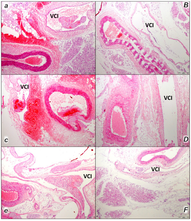
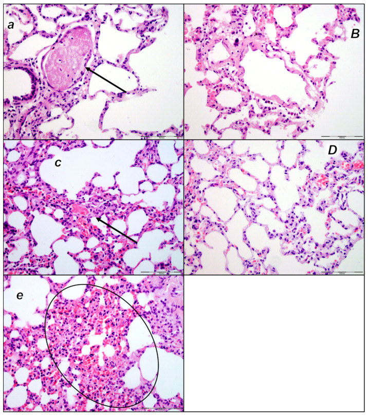
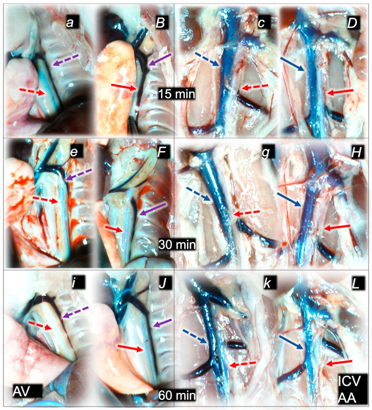
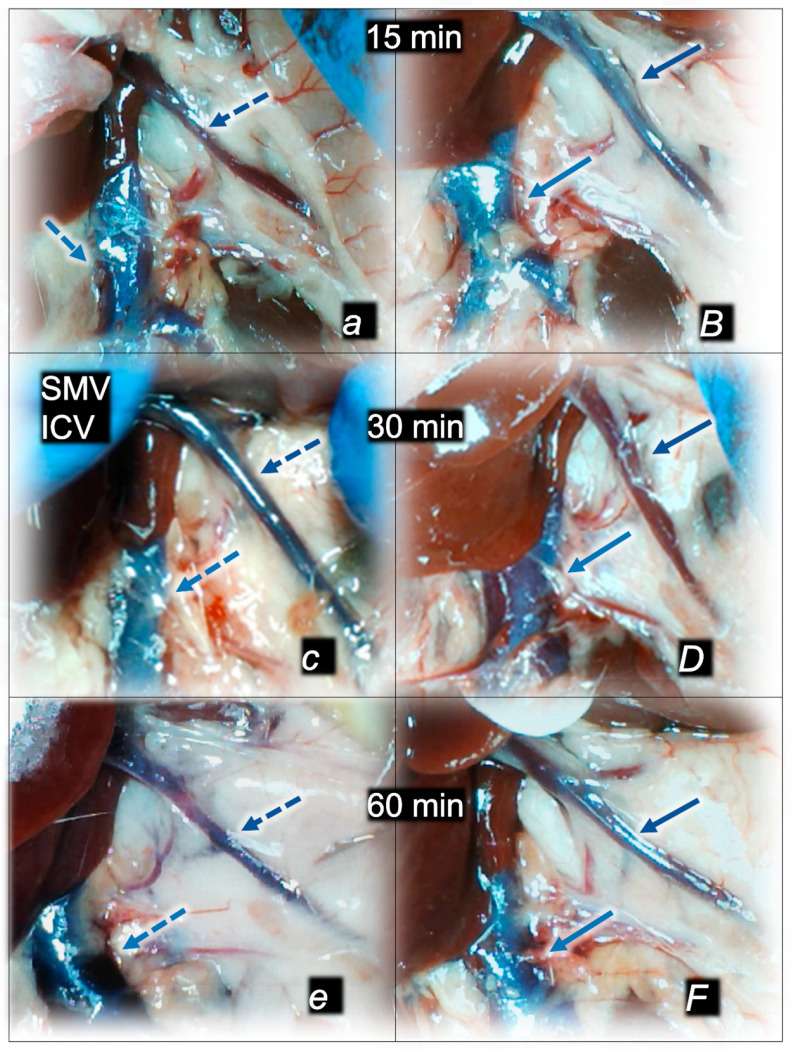
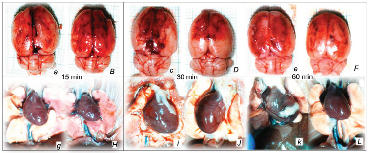
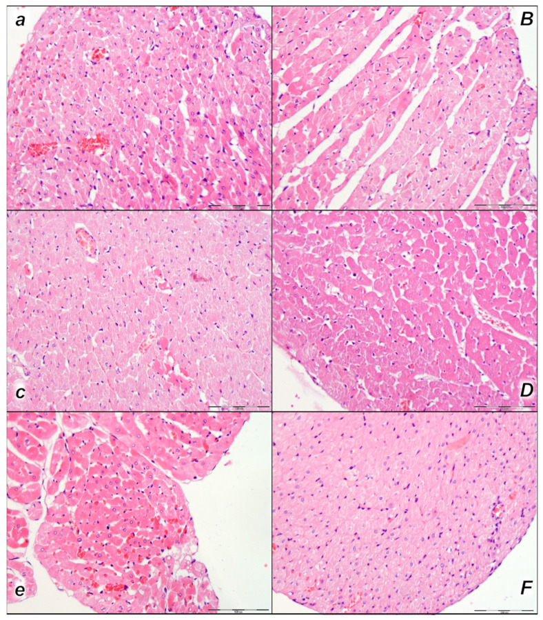
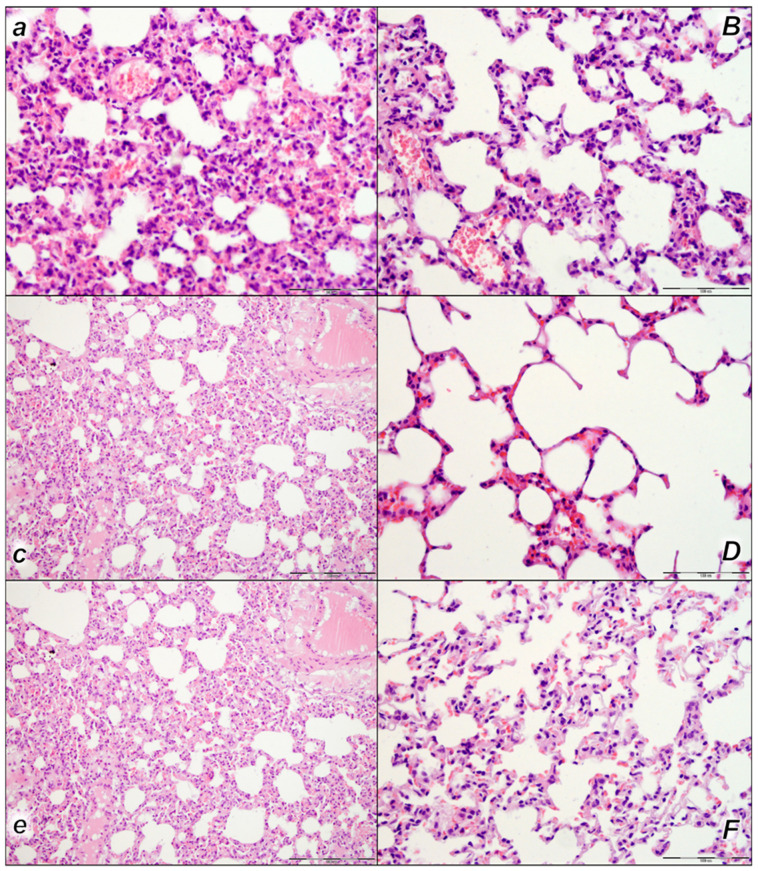
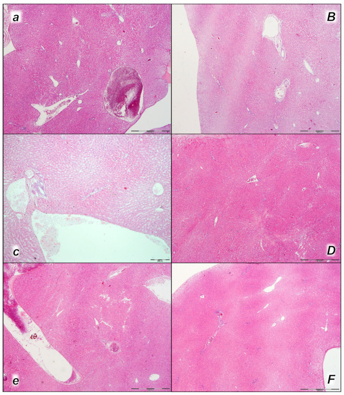
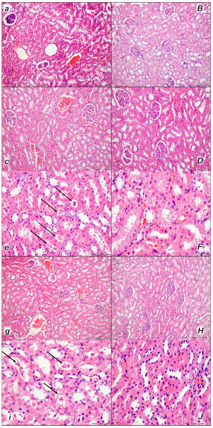
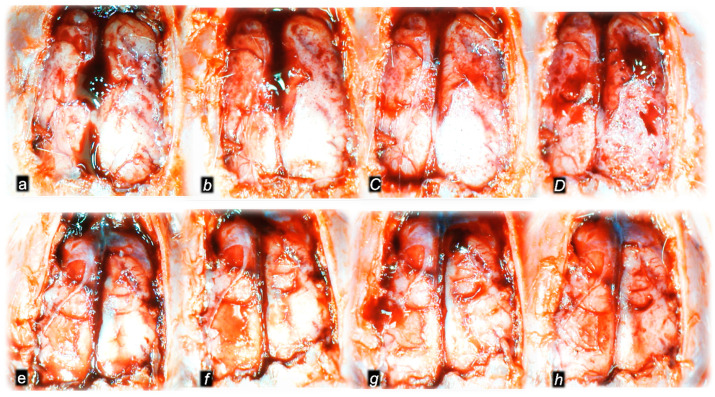
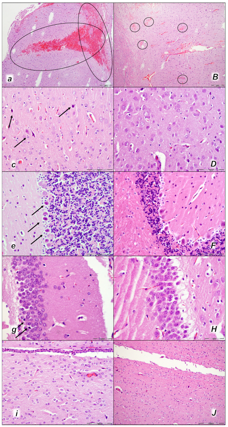
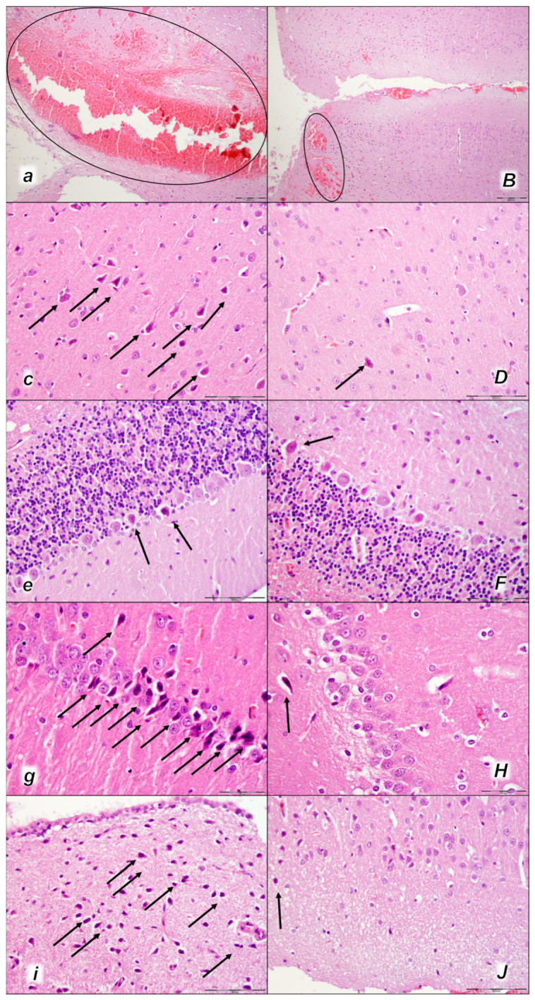
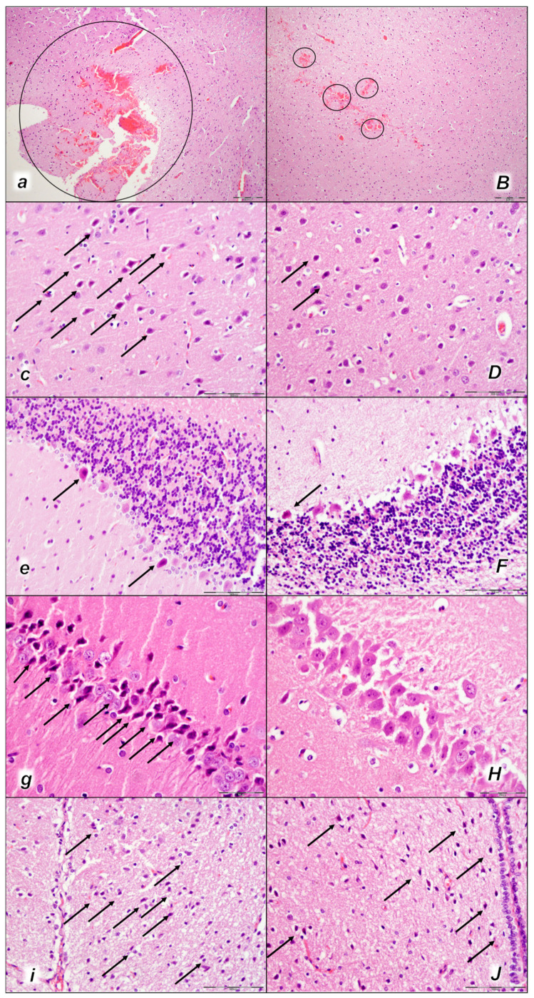
After inferior caval vein embolization therapy, post-embolization syndrome (sodium laurate 10 mg/kg, 0.1 mL into rat inferior caval vein, assessment at 15, 30, 60 min, prime lung lesions, thromboemboli occluding lung vessels), as a severe occlusion/occlusion-like syndrome, might be resolved as a whole by stable gastric pentadecapeptide BPC 157 therapy. At 5 min after laurate injection, stable gastric pentadecapeptide BPC 157 was implemented as therapy (10 µg/kg, 10 ng/kg intraperitoneally or intragastrically). As before, confronted with the occlusion of major vessel(s) or similar noxious procedures, such as rapidly acting Virchow triad circumstances, the particular effect of the therapy (i.e., collateral pathways activation, "bypassing vascular key", i.e., direct blood flow delivery via activation of azygos vein) assisted in the recovery of the vessel/s and counteracted multiorgan failure due to occlusion/occlusion-like syndrome as a whole in the laurate-injected rats. Along with prime lung lesions and thromboemboli occluding lung vessels, post-embolization syndrome rapidly occurred peripherally and centrally as a shared multiorgan and vessel failure, brain, heart, lung, liver, kidney, and gastrointestinal tract lesions, venous hypertension (intracranial (superior sagittal sinus), portal, and caval), aortal hypotension, progressing thrombosis in veins and arteries and stasis, congested and/or failed major veins, and severe ECG disturbances. Whatever the cause, these were all counteracted, eliminated, or attenuated by the application of BPC 157 therapy. As recovery with BPC 157 therapy commonly and rapidly occurred, reversing the collapsed azygos vein to the rescuing collateral pathway might initiate rapid direct blood delivery and start blood flow reorganization. In conclusion, we suggest BPC 157 therapy to resolve further vascular and embolization injuries.

Keywords: general occlusion/occlusion-like syndrome; post-embolization syndrome; rats; stable gastric pentadecapeptide BPC 157; therapy.


Conflict of interest statement

The authors declare no conflict of interest.

Figures

Figure 1
Figure 1
Thromboemboli presentation in vessels (aF). In control rats, vascular thromboemboli presented within the lumen of large retroperitoneal vessels, abdominal aorta, and inferior vena cava (VCI) in all the time periods (15 min (a), 30 min (c), 60 min (e)) following laurate application into inferior caval vein. Thrombi consisted of fibrin and blood cells. In contrast, no thrombi were observed in treated animals. No thromboemboli were observed in BPC 157-treated group (15 min (B), 30 min (D), 60 min (F). (HE staining; magnification 100×; scale bar 200 μm).
Figure 2
Figure 2
Thromboemboli presentation in lung (ae). In control rats (small italic letters), thromboemboli (black arrow) were found within the lumen of medium and small lung and vascular vessels causing luminal occlusion, at two assessment time periods (30 min (a) and 60 min (c) following laurate application into inferior caval vein), with regional intraalveolar hemorrhagia (marked area) of lung parenchyma following thromboembolism after the time period of 60 min (e). Thromboemboli were absent in BPC 157-treated rats (capital italic letters) (30 min (B) and 60 min (D) following laurate application into the inferior caval vein. (HE staining; magnification 400×; scale bar 100 μm).
Figure 3
Figure 3
Illustrative presentation of azygos vein (AV) (violet arrows) and aorta (AA) in abdomen or thorax (red arrows) and inferior caval vein (blue arrows) (ICV) (aL) in rats that received saline (control, small italic letters, dashed arrows) or BPC 157 therapy (capital italic letters, full arrows), immediately before sacrifice. Azygos vein and aorta at 15 min (a,B), 30 min (c,D), and 60 min (e,F) following application of 0.1 mL of sodium laurate (10 mg/kg) into the inferior caval vein. Inferior caval vein and abdominal aorta at 15 min (c,D), 30 min (g,H), and 60 min (k,H) following application of 0.1 mL of sodium laurate (10 mg/kg) into the inferior caval vein.
Figure 4
Figure 4
Illustrative presentation of superior mesenteric vein (SMV, dark blue arrows) and inferior caval vein (ICV, light blue arrows) (a,B,c,D,e,F) in rats that received 0.1 mL of sodium laurate (10 mg/kg) into the inferior caval vein, and then saline (control, small italic letters, dashed blue arrows) or BPC 157 therapy (capital italic letter, full blue arrows), immediately before sacrifice. Presentation at 15 min (a,B), 30 min (c,D), and 60 min (e,F) following application of 0.1 mL of sodium laurate (10 mg/kg) into the inferior caval vein.
Figure 5
Figure 5
Illustrative presentation of brain (swelling) (a,B,c,D,e,F) and heart (dilatation) (g,H,i,J,k,L) in rats that in rats that received 0.1 mL of sodium laurate (10 mg/kg) into the inferior caval vein, and then saline (control, small italic letters) or BPC 157 therapy (capital italic letters). Presentation immediately after sacrifice (brain) or immediately before sacrifice (heart) at 15 min (a,B,g,H), 30 min (c,D,i,J) and 60 min (e,F,k,L) following application of 0.1 mL of sodium laurate (10 mg/kg) into the inferior caval vein.
Figure 6
Figure 6
Heart, microscopy presentation (aF). Pronounced congestion and dilatation of coronary arteries and their intramyocardial branches up to the subendocardial area in control rats (small italic letters) at 15 min (a), 30 min (c), and 60 min (e) after laurate application into inferior caval vein. No changes in BPC 157-treated rats (capital italic letters) were observed at 15 min (B), 30 min (D), and 60 min (F) after laurate application into the inferior caval vein. (HE staining; magnification 400×; scale bar 100 μm).
Figure 7
Figure 7
Lung, microscopy presentation (aF). In the control rats, within-lung parenchyma thickening of the alveolar membranes was observed due to capillary congestion, pulmonary edema, and dilatation of larger blood vessels (small italic letters) at 15 min (a), 30 min (c), and 60 min (e) after laurate application into the inferior caval vein. No changes in BPC 157-treated rats were observed (capital italic letters) at 15 min (B), 30 min (D), and 60 min (F) after laurate application into the inferior caval vein. (HE staining; magnification 400×; scale bar 100 μm).
Figure 8
Figure 8
Liver, microscopy presentation (aF). In liver parenchyma, pronounced dilatation of sinusoids and branches of the portal vein in portal tracts was found in liver tissue of control rats (small italic letters) at 15 min (a), 30 min (c), and 60 min (e) after laurate application into the inferior caval vein. No changes in BPC 157-treated rats (capital italic letters) were observed at 15 min (B), 30 min (D), and 60 min (F) after laurate application into the inferior caval vein. (HE staining; magnification 400×; scale bar 100 μm).
Figure 9
Figure 9
Kidney, microscopy presentation (aF). After laurate application into the inferior caval vein control rats (small italic letters), moderate vascular congestion was shown in renal parenchyma, interstitial edema at 15 min (a), and moderate vascular congestion, and interstitial edema, and intratubular hyaline casts (black arrows) at 30 min (c,e) and at 60 min (g,i). No changes in BPC 157-treated rats (capital italic letters) were observed at 15 min (B), 30 min (D,F), and 60 min (H,J) after laurate application into the inferior caval vein. (HE staining; magnification 200×; scale bar 200 μm (a,B,c,D,g,H); magnification 600×; scale bar 50 μm (e,F,i,J)).
Figure 10
Figure 10
Gross brain presentation in healthy rats (normal small letters) and then after laureate application into inferior caval vein (italic letters), and subsequent application of the BPC 157 therapy (capital italic letters) or saline (small italic letters) (ah). Brain presentation in normal healthy rats (a,e), brain swelling presentation immediately upon laurate application (b,f). Then, there was the opposite effect of therapy: decreased brain swelling immediately upon BPC 157 administration (C), decreased brain swelling in BPC 157-treated rats immediately before sacrifice (D), in contrast to further brain swelling immediately upon saline administration (g) and increased brain swelling in saline-treated rats immediately before sacrifice (h). A similar presentation was noted with both intragastric and intraperitoneal BPC 157 therapy.
Figure 11
Figure 11
Brain neuropathological changes, 15 min following laurate application into the inferior caval vein, (aJ). In the control rats (small italic letters), a pronounced edema and congestion in the brain tissue were observed. Focal, pronounced, and deep intracerebral hemorrhage involving areas of brain tissue was observed, affecting areas of the neocortex, the corpus callosum, the amygdala, and the striatum in the brain tissue (a) (marked area). Mild neurodegenerative changes of the central nervous system, such as rare karyopyknotic cells affecting cerebral (c) and cerebellar (e) cortex, a karyopyknosis and degeneration of Purkinje cells of the cerebellar cortex, and karyopyknosis of cortical neurons, were observed (black arrows), including in the hypothalamus/thalamus (g) and hippocampus (i). In the BPC 157-treated rats (capital italic letters), mild edema and congestion in the brain tissue. Intracerebral hemorrhage was visible only within superficial layers of the neocortex (marked area) (B). No neurodegenerative changes of the central nervous system were observed in BPC 157 rats: in the cerebrum (D), cerebellum (F), hypothalamus/thalamus (H), and hippocampus (J). (HE staining; magnification 100×; scale bar 200 µm (a,B); magnification 400×; scale bar 100 μm (cJ)).
Figure 12
Figure 12
Brain neuropathological changes 30 min following laurate application into the inferior caval vein (aJ). In the control rats (italic small letters), a pronounced edema and congestion in the brain tissue were observed. Focal, pronounced, and deep intracerebral hemorrhage involving areas of brain tissue was observed, affecting areas of the neocortex, the corpus callosum, the amygdala, and the striatum in the brain tissue (a) (marked area). Moderate and severe neurodegenerative changes of the central nervous system, such as rare karyopyknotic cells affecting the cerebral (c) and cerebellar (e) cortex, a karyopyknosis and degeneration of the Purkinje cells of the cerebellar cortex, and karyopyknosis of cortical neurons, were observed (black arrows), as well as karyopyknosis of cortical neurons, hypothalamic neurons (g), and pyramidal cells of the hippocampus (i) (black arrows). In the BPC 157-treated rats (capital italic letters), only mild edema and congestion in the brain tissue were observed. Intracerebral hemorrhage was visible only within superficial layers of the neocortex (marked area) (B). BPC 157-treated rats presented no or only rare karyopyknotic cells in all four regions: the cerebrum (D), cerebellum (F), hypothalamus/thalamus (H), and hippocampus (J). (HE staining; magnification 100×; scale bar 200 µm (a,B); magnification 400×; scale bar 100 µm (cJ)).
Figure 13
Figure 13
Brain neuropathological changes 60 min following laurate application into the inferior caval vein (aJ). In the control rats (small italic letters), a pronounced edema and congestion in the brain tissue were observed. Focal, pronounced, and deep intracerebral hemorrhage involving areas of brain tissue was observed, affecting areas of the neocortex, the corpus callosum, the amygdala, and the striatum in the brain tissue (a) (marked area). Moderate and severe neurodegenerative changes of the central nervous system, such as rare karyopyknotic cells affecting the cerebral (c) and cerebellar (e) cortex, a karyopyknosis and degeneration of the Purkinje cells of the cerebellar cortex, and karyopyknosis of cortical neurons, were observed (black arrows), as well as karyopyknosis of cortical neurons, hypothalamic neurons (g), and pyramidal cells of the hippocampus (i) (black arrows). In the BPC 157-treated rats (capital italic letters), mild edema and congestion in the brain tissue were observed. Intracerebral hemorrhage was visible only within superficial layers of the neocortex (marked area) (B). BPC 157-treated rats presented no or only rare karyopyknotic cells in all four regions: the cerebrum (D), cerebellum (F), hypothalamus/thalamus (H), and hippocampus (J). ((HE staining; magnification 100×; scale bar 200 µm (a,B); magnification 400×; scale bar 100 µm (cJ)).

Similar articles

Cited by

References

    1. Vukojevic J., Milavic M., Perovic D., Ilic S., Zemba Cilic A., Duran N., Strbe S., Zoricic Z., Filipcic I., Brecic P., et al. Pentadecapeptide BPC 157 and the central nervous system. Neural Regen. Res. 2022;17:482–487. doi: 10.4103/1673-5374.320969. - DOI - PMC - PubMed
    1. Seiwerth S., Milavic M., Vukojevic J., Gojkovic S., Krezic I., Vuletic L.B., Pavlov K.H., Petrovic A., Sikiric S., Vranes H., et al. Stable gastric pentadecapeptide BPC 157 and wound healing. Front. Pharmacol. 2021;12:627533. doi: 10.3389/fphar.2021.627533. - DOI - PMC - PubMed
    1. Sikiric P., Hahm K.B., Blagaic A.B., Tvrdeic A., Pavlov K.H., Petrovic A., Kokot A., Gojkovic S., Krezic I., Drmic D., et al. Stable gastric pentadecapeptide BPC 157, Robert’s stomach cytoprotection/adaptive cytoprotection/organoprotection, and Selye’s stress coping response: Progress, achievements, and the future. Gut Liver. 2020;14:153–167. doi: 10.5009/gnl18490. - DOI - PMC - PubMed
    1. Park J.M., Lee H.J., Sikiric P., Hahm K.B. BPC 157 rescued NSAID-cytotoxicity via stabilizing intestinal permeability and enhancing cytoprotection. Curr. Pharm. Des. 2020;26:2971–2981. doi: 10.2174/1381612826666200523180301. - DOI - PubMed
    1. Sikiric P., Rucman R., Turkovic B., Sever M., Klicek R., Radic B., Drmic D., Stupnisek M., Misic M., Vuletic L.B., et al. Novel cytoprotective mediator, stable gastric pentadecapeptide BPC 157. Vascular recruitment and gastrointestinal tract healing. Curr. Pharm. Des. 2018;24:1990–2001. doi: 10.2174/1381612824666180608101119. - DOI - PubMed