. 2023 Oct 11;15(10):2447.
doi: 10.3390/pharmaceutics15102447.

Anticancer Peptides Derived from Aldolase A and Induced Tumor-Suppressing Cells Inhibit Pancreatic Ductal Adenocarcinoma Cells

Affiliations

Anticancer Peptides Derived from Aldolase A and Induced Tumor-Suppressing Cells Inhibit Pancreatic Ductal Adenocarcinoma Cells

Changpeng Cui et al. Pharmaceutics. .

Abstract

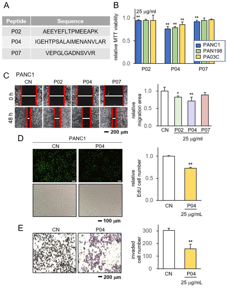
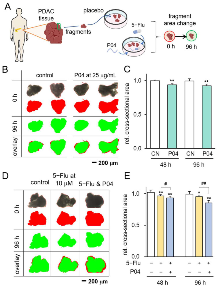
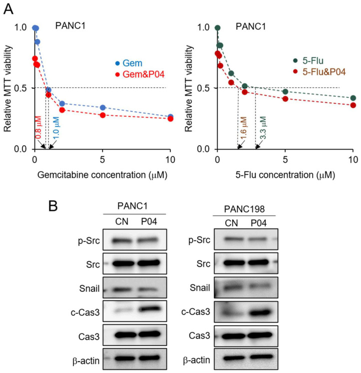
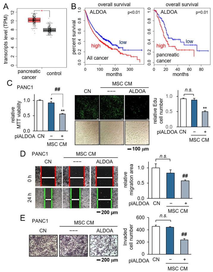
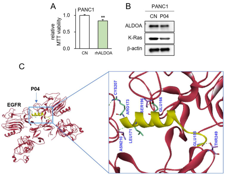
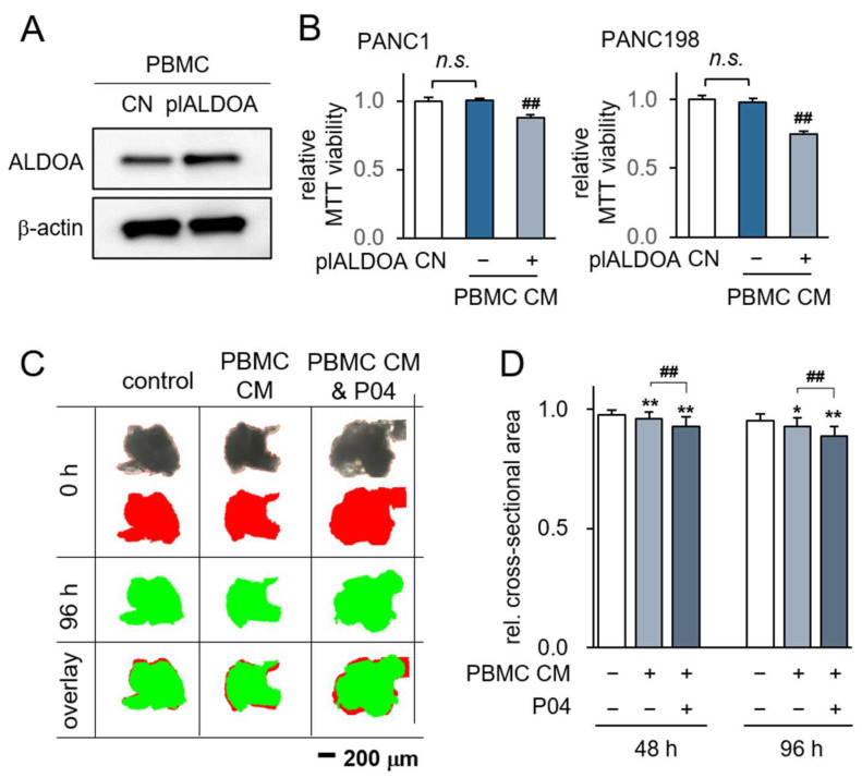
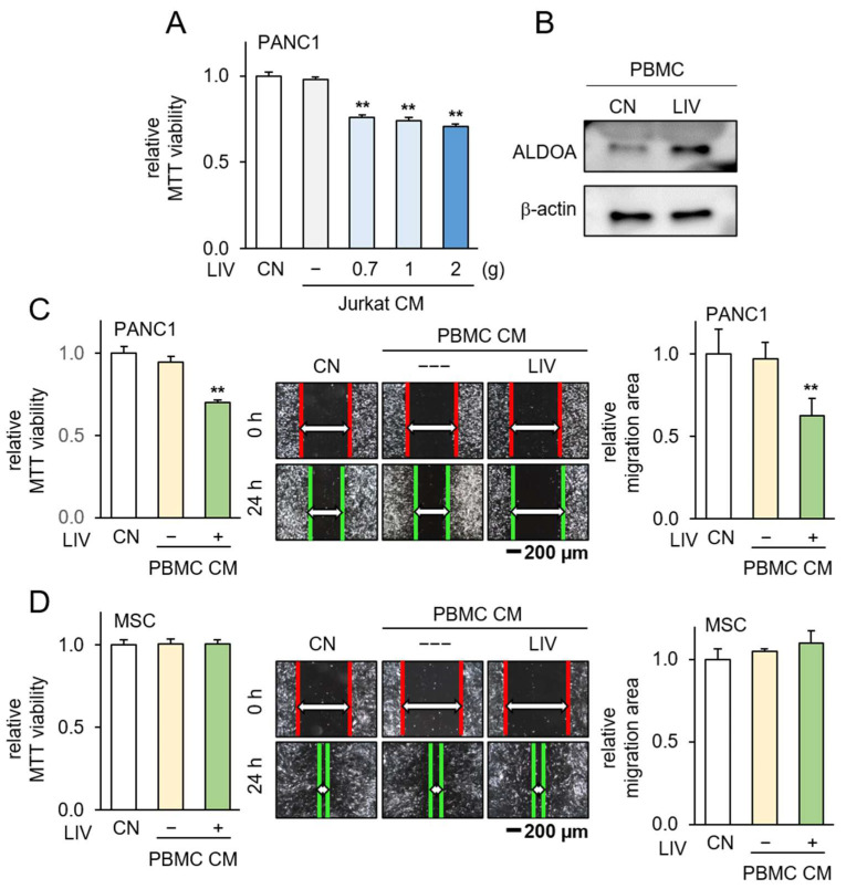
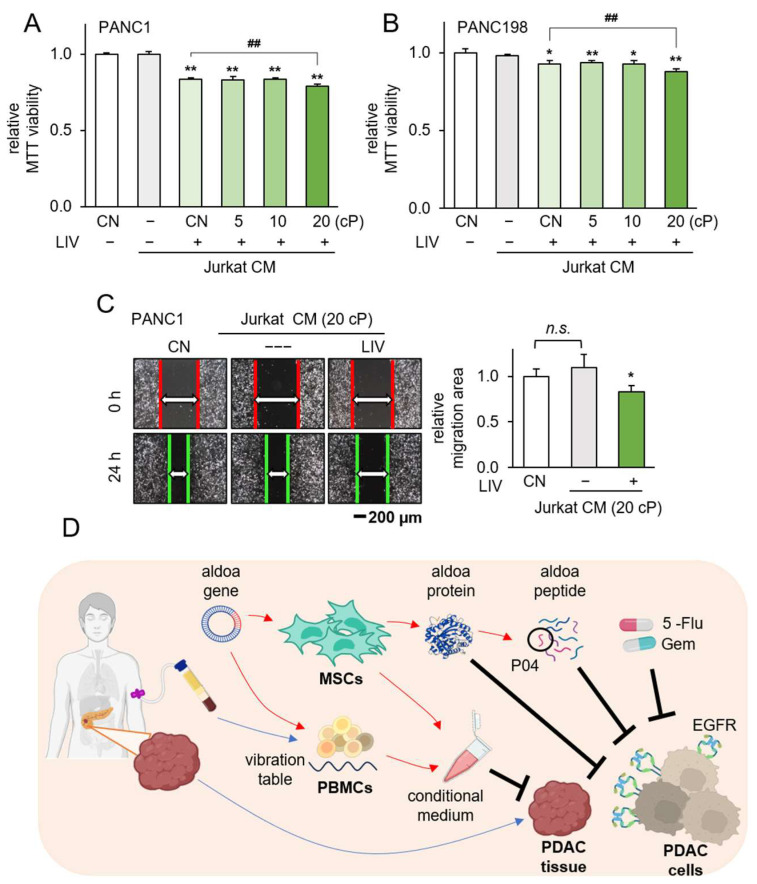
PDAC (pancreatic ductal adenocarcinoma) is a highly aggressive malignant tumor. We have previously developed induced tumor-suppressing cells (iTSCs) that secrete a group of tumor-suppressing proteins. Here, we examined a unique procedure to identify anticancer peptides (ACPs), using trypsin-digested iTSCs-derived protein fragments. Among the 10 ACP candidates, P04 (IGEHTPSALAIMENANVLAR) presented the most efficient anti-PDAC activities. P04 was derived from aldolase A (ALDOA), a glycolytic enzyme. Extracellular ALDOA, as well as P04, was predicted to interact with epidermal growth factor receptor (EGFR), and P04 downregulated oncoproteins such as Snail and Src. Importantly, P04 has no inhibitory effect on mesenchymal stem cells (MSCs). We also generated iTSCs by overexpressing ALDOA in MSCs and peripheral blood mononuclear cells (PBMCs). iTSC-derived conditioned medium (CM) inhibited the progression of PDAC cells as well as PDAC tissue fragments. The inhibitory effect of P04 was additive to that of CM and chemotherapeutic drugs such as 5-Flu and gemcitabine. Notably, applying mechanical vibration to PBMCs elevated ALDOA and converted PBMCs into iTSCs. Collectively, this study presented a unique procedure for selecting anticancer P04 from ALDOA in an iTSCs-derived proteome for the treatment of PDAC.

Keywords: ALDOA; induced tumor-suppressing cells; pancreatic ductal carcinoma; peptide.


Conflict of interest statement

The authors declare no conflict of interest.

Figures

Figure 1
Figure 1
Anti-tumor effect of P04 on PDAC cell lines. CN = control. The single and double asterisks indicate p < 0.05 and 0.01, respectively. (A) The amino acid sequences of the selected three peptides, including P02, P04, and P07; (B) reduction in MTT-based viability of PANC1, PAN198, and PA03C pancreatic tumor cells in response to 25 μg/mL of P02, P04, and P07; (C) reduction in scratch-based motility of PANC1 cells in response to 25 μg/mL of P04; (D,E) suppression of EdU-based proliferation and transwell-based invasion of PANC1 cells in response to 25 μg of P04, respectively.
Figure 2
Figure 2
Inhibitory effects on PDAC tissue fragments in response to P04. The single and double asterisks indicate p < 0.05 and 0.01 vs. CN, respectively. The single and double hashtag indicate p < 0.05 and 0.01, 5-Flu group vs. 5-Flu&P04 group, respectively (A) The illustration of PDAC tissue fragments assay; (B,C) reduction in PDAC fragment size in 96 h in response to 25 μg/mL of P04 (n = 8). The red image indicates tissue fragments at 0 h, while the green image indicates them at 96 h; (D,E) reduction in PDAC fragment size in 96 h in response to 10 μM of 5-Flu and the combination with 25 μg/mL P04 is more effective (n = 8). The red image indicates tissue fragments at 0 h, which represents the area of the black original image, while the green image indicates them at 96 h.
Figure 3
Figure 3
Inhibitory effects of P04 in combination with gemcitabine and 5-Flu. CN = control, Gem = gemcitabine, and 5-Flu = 5-fluorouracil. (A) Additive anti-tumor effects of P04, together with gemcitabine or 5-Flu; (B) decrease in the levels of p-Src and Snail, as well as an increase in cleaved caspase 3 (c-Cas-3) in PANC1 and PAN198 PDAC cells in response to 25 μg/mL of P04.
Figure 4
Figure 4
Generation of iTSCs by the overexpression of ALDOA in MSCs. CN = control, pl = plasmid transfection, MSC = mesenchymal stem cell, and CM = conditioned medium. The single and double asterisks indicate p < 0.05 and 0.01, MSC CM group vs. CN. The double hashtag indicates p < 0.01, MSC-plALDOA CM group vs. MSC-NC CM group, respectively. n.s. = non-significant. (A,B) The elevated ALDOA transcript level in patients with PDAC, and lower survival rate for patients with a high ALDOA level in TCGA database; (CE) reduction in MTT-based viability, EdU-based proliferation, scratch-based motility, and transwell invasion, respectively, by ALDOA-overexpressing MSC-derived conditioned medium.
Figure 5
Figure 5
Predicted interaction between P04 and EGFR. The double asterisk indicates p < 0.01 vs. CN (A) Reduction in MTT-based viability of PANC1 cells in response to 5 μg/mL of recombinant ALDOA protein; (B) decrease in the levels of ALDOA and K-Ras in PANC1 cells by the application of 25 μg/mL of P04; (C) predicted interactions between P04 and EGFR by molecular docking using Z-DOCK software 2016.
Figure 6
Figure 6
Generation of iTSCs from PBMCs by overexpressing ALDOA. CN = control, pl = plasmid transfection, PBMC = peripheral blood mononuclear cells, and CM = conditioned medium. (A) Elevated ALDOA level by transfection of ALDOA plasmids; (B) reduction in MTT-based viability of PANC1 and PAN198 cells by ALDOA-overexpressing PBMC-derived CM. The double hashtag indicates p < 0.01, PBMC-plALDOA CM group vs. PBMC-NC CM group, respectively. n.s. = non-significant; (C,D) shrinkage of PDAC fragments by ALDOA-overexpressing PBMC-derived CM with and without P04. The red image indicates tissue fragments at 0 h, which represents the area of the black original image, while the green image indicates them at 96 h. The single and double asterisk indicates p < 0.05 and 0.01 vs. CN, respectively. The double hashtag indicates p < 0.01, PBMC CM & P04 group vs. PBMC CM group.
Figure 7
Figure 7
Generation of iTSCs from PBMCs by the application of low-intensity vibration. CM = conditioned medium, CN = control, and LIV = vertical low-intensity vibration. The double asterisk indicates p < 0.01 vs. non-LIV treated group. PBMC CM & P04 group vs. PBMC CM group. (A) Reduction in MTT-based viability of PANC1 cells by the application of LIV-treated Jurkat cell-derived CM; (B) an elevated level of ALDOA in PBMCs in response to LIV; (C) reduction in MTT-based viability and scratch-based motility of PANC1 cells by LIV-treated PBMC-derived CM; (D) no detectable changes in MTT-based viability and scratch-based motility of MSCs by LIV-treated PBMC-derived CM.
Figure 8
Figure 8
Generation of iTSCs from Jurkat cells by LIV at different viscosity. CM = conditioned medium, CN = control, LIV = vertical low-intensity vibration, and cP = centipoise (viscosity unit). (A,B) Reduction in MTT-based viability of PANC1 and PAN198 cells by the application of LIV-treated Jurkat-derived CM. The single and double asterisk indicates p < 0.05 and 0.01, respectively vs. non-LIV treated Jurkat CM group. The double hashtag indicates p < 0.01, Jurkat LIV treated at 20 cP CM group & Jurkat LIV treated at normal medium CM group. n.s. = non-significant; (C) reduction in scratch-based motility of PANC1 cells by LIV-treated Jurkat-derived CM; (D) proposed mechanism of the anti-tumor action of ALDOA, P04, and vibration-treated PBMC-derived CM.

References

    1. Moffat G.T., Epstein A.S., O’Reilly E.M. Pancreatic cancer-A disease in need: Optimizing and integrating supportive care. Cancer. 2019;125:3927–3935. doi: 10.1002/cncr.32423. - DOI - PMC - PubMed
    1. Sturm N., Ettrich T.J., Perkhofer L. The Impact of Biomarkers in Pancreatic Ductal Adenocarcinoma on Diagnosis, Surveillance and Therapy. Cancers. 2022;14:217. doi: 10.3390/cancers14010217. - DOI - PMC - PubMed
    1. Liu Z., Hayashi H., Matsumura K., Uemura N., Shiraishi Y., Sato H., Baba H. Biological and Clinical Impacts of Glucose Metabolism in Pancreatic Ductal Adenocarcinoma. Cancers. 2023;15:498. doi: 10.3390/cancers15020498. - DOI - PMC - PubMed
    1. Nagai M., Sho M., Akahori T., Nakagawa K., Nakamura K. Application of liquid biopsy for surgical management of pancreatic cancer. Ann. Gastroenterol. Surg. 2020;4:216–223. doi: 10.1002/ags3.12317. - DOI - PMC - PubMed
    1. Schwarz L., Vernerey D., Bachet J.B., Tuech J.J., Portales F., Michel P., Cunha A.S. Resectable pancreatic adenocarcinoma neo-adjuvant FOLF(IRIN)OX-based chemotherapy—A multicenter, non-comparative, randomized, phase II trial (PANACHE01-PRODIGE48 study) BMC Cancer. 2018;18:762. doi: 10.1186/s12885-018-4663-4. - DOI - PMC - PubMed