Review
. 2023 Oct 12;15(20):4059.
doi: 10.3390/polym15204059.

The Progress in the Application of Dissolving Microneedles in Biomedicine

Affiliations
Review

The Progress in the Application of Dissolving Microneedles in Biomedicine

Xueqing Yu et al. Polymers (Basel). .

Abstract

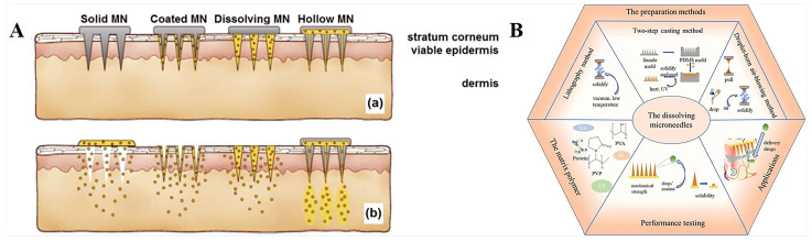
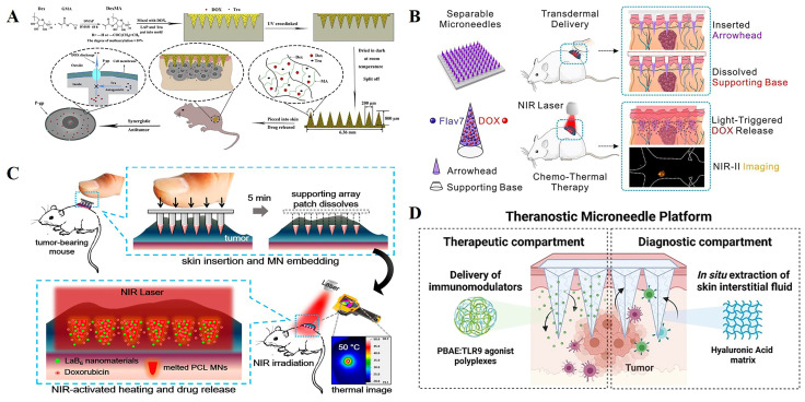
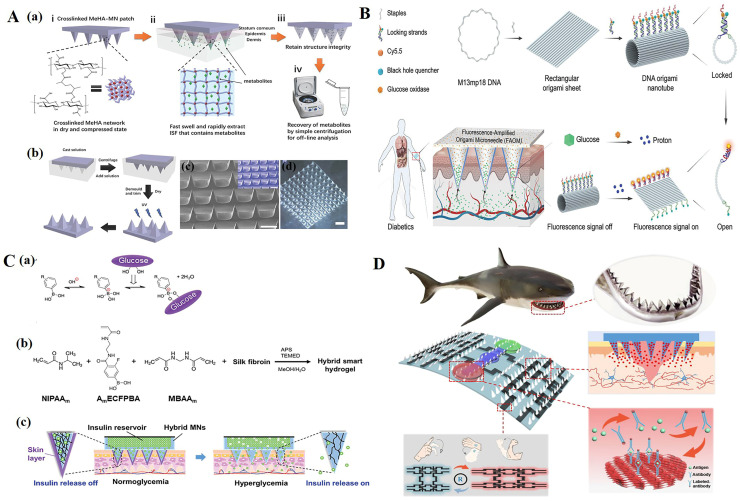
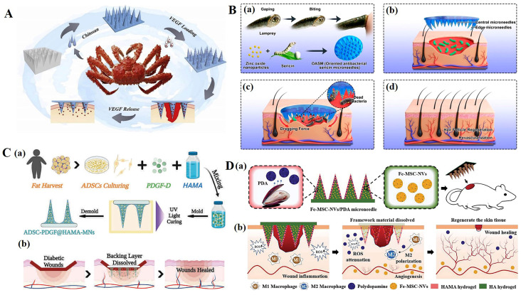
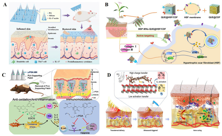
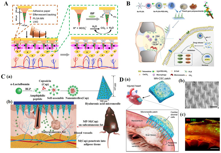
In recent years, microneedle technology has been widely used for the transdermal delivery of substances, showing improvements in drug delivery effects with the advantages of minimally invasive, painless, and convenient operation. With the development of nano- and electrochemical technology, different types of microneedles are increasingly being used in other biomedical fields. Recent research progress shows that dissolving microneedles have achieved remarkable results in the fields of dermatological treatment, disease diagnosis and monitoring, and vaccine delivery, and they have a wide range of application prospects in various biomedical fields, showing their great potential as a form of clinical treatment. This review mainly focuses on dissolving microneedles, summarizing the latest research progress in various biomedical fields, providing inspiration for the subsequent intelligent and commercial development of dissolving microneedles, and providing better solutions for clinical treatment.

Keywords: cancer therapy; cutaneous disease; diagnostic; dissolving microneedles; drug delivery; wound healing.


Conflict of interest statement

The authors declare no conflict of interest.

Figures

Figure 1
Figure 1
(A). Schematic diagram of different types of microneedles applied to (a) skin and (b) drug delivery [23]. (B). Relevant content of dissolution microneedles [28]. Reproduced with permission from the Journal of Controlled Release and International Journal of Pharmaceutics, respectively.
Figure 2
Figure 2
(A). Schematic of DexMA hydrogel MNs that can be used for continuous transdermal release of drugs [43]. (B). Flav7 + DOX co-loaded separable MNs for light-triggered chemo-thermal therapy of SMTs [45]. (C). Schematic illustrations of a combination of chemotherapy and photothermal therapy using near-infrared (NIR) light-activatable microneedles (MNs) [46]. (D). Representative scheme of a hyaluronic acid (HA)-based microneedle platform for the delivery of immunomodulatory drugs (CpG-ODNs), complexed with poly (beta-amino esters) (PBAEs) and simultaneous sampling of interstitial fluid (ISF) for the recovery of immune cells ex vivo [53]. Reproduced with permission from the Carbohydrate Polymers, Chemical Engineering Journal, ACS Nano, and Theranostics, respectively.
Figure 4
Figure 4
(A) (a) Schematic representation of the rapid extraction of ISF by crosslinked MeHA-MN patches. (i) The crosslinked MeHA network within MN patch is dried and compressed. (ii) In skin, the MeHA-MN patch rapidly swells and extracts ISF that contains metabolites. (iii) After removal, MN patch retains the structure integrity. (iv) Extracted ISF and metabolites can be efficiently recovered from MN patch by centrifugation for subsequent offline analysis. (b) Schematic of the fabrication process of a crosslinked MeHA-MN patch. (c) Scanning electron microscopy (SEM) image of a crosslinked MeHA-MN patch; inlet is a false-color image (scale bar: 500 µm). (d) Optical image of a crosslinked MeHA-MN patch (scale bar: 1000 µm) [92]. (B) Schematic illustration of a fluorescence-amplified origami microneedle (FAOM) device for quantitatively monitoring blood glucose [97]. (C) (a) Glucose-dependent equilibria of PBA derivatives. (b) Schematic representation of the formation of semi-IPN hydrogel. (c) “Skin-layer” controlled glucose-responsive insulin release from the MN array patch [98]. (D) Schematic diagram of shark-tooth-inspired microneedle dressing for intelligent wound management including motion sensing, biochemical analysis, and healing [112]. Reproduced with permission from Advanced Materials, Advanced Functional Materials, and Acs Nano, respectively.
Figure 3
Figure 3
(A). Scheme of the fabrication and controllable drug release application of the biomass microneedle patch [70]. (B). Design and fabrication of bioinspired oriented antibacterial sericin microneedles (OASM) for healing infected wounds. (a) Lamprey-teeth-inspired OASM was composed of sericin and zinc oxide nanoparticles (ZNPs), which would degrade and release ZNPs into the wounds. (b) The central short needles would insert into the wound site, and the edged long needles possessed a tilt angle that would provide a dragging force. (c) OASM would penetrate the skin tissues of the infected wound, the released ZNPs could kill the bacteria, and the edged titled needles would provide a dragging force to stimulate wound contraction. (d) Hair follicle regeneration and revascularization can be observed in the wound after OASM treatment. [71]. (C). (a) Schematic illustrations of the MN system loaded with ADSCs and PDGF-D. (b) Application of MNs for diabetic wound treatment [77]. (D). Schematic illustrations of the Fe-MSC-NVs/PDA MN patch for diabetic wound healing: (a) schematic of Fe-MSC-NVs/PDA MN patch; (b) schematic of the wound closure process [78]. Reproduced with permission from Bioactive Materials, Nano Letters, Advanced Functional Materials, and Advanced Science, respectively.
Figure 5
Figure 5
(A) Schematic design of the application of mAbs-loaded photothermal responsive MN patch for psoriasis treatment [135]. (B) Schematic illustration of fabrication and administration of BSP-MNs-QUE@HSF/CDF [136]. (C) Schematic illustration of the use of epigallocatechin gallate (EGCG)/L-ascorbic acid (AA)-loaded poly-γ-glutamate (γ -PGA) microneedles (MNs) to ameliorate AD-like symptoms in mice [137]. (D) Sonocatalytic mechanism and the treatment of acne through efficient sonodynamic ion therapy–based MN patch. US: ultrasound [138]. Reproduced with permission from Advanced Functional Materials, ACS Nano, Acta Biomaterialia, and Science Advances, respectively.
Figure 6
Figure 6
(A) Long-acting, reversible contraception using an effervescent microneedle patch [37]. (B) Drug-loaded multifunctional PLGA nanoparticles integrated with GP-fabricated dissolving microneedles for the local treatment of RA [144]. (C) Schematic illustration of a multifunctional microneedle patch with capsaicin-loaded micelles for suppressing adipogenesis and promoting adipocyte browning. (a) Micelles encapsulated with capsaicin (M (Cap)) are prepared from self-assembly of α-lac peptides and Cap; M (Cap) were then loaded into the microneedle patch made of hyaluronic acid (HA) and polyvinyl alcohol (PVA) with body temperature responsive melting property. (b) The microneedle patch (MP) encapsulated with M (Cap) was pressed onto the fur-removed skin of the left abdominal subcutaneous fat of high fat diet (HFD)-induced obese mice. [149] (D). Characterization of MN-CSCs. (a) Schematic showing the overall design used to test the therapeutic benefits of MN-CSCs for infarcted hearts. (b) SEM image of MN (scale bar: 500 μm). (c) Representative fluorescent image indicating that DiO-labeled CSCs (green) were encapsulated in fibrin gel and then integrated onto the top surface of the MN array (red) (scale bar: 500 μm) [161]. Reproduced with permission from Science Advances, Chemical Engineering Journal, Advanced Functional Materials, and Advanced Healthcare Materials, respectively.

Similar articles

Cited by

References

    1. Allen E.A., O'Mahony C., Cronin M., O'Mahony T., Moore A.C., Crean A.M. Dissolvable microneedle fabrication using piezoelectric dispensing technology. Int. J. Pharm. 2016;500:1–10. doi: 10.1016/j.ijpharm.2015.12.052. - DOI - PubMed
    1. Badran M.M., Kuntsche J., Fahr A. Skin penetration enhancement by a microneedle device (Dermaroller) in vitro: Dependency on needle size and applied formulation. Eur. J. Pharm. Sci. 2009;36:511–523. doi: 10.1016/j.ejps.2008.12.008. - DOI - PubMed
    1. Engelke L., Winter G., Hook S., Engert J. Recent insights into cutaneous immunization: How to vaccinate via the skin. Vaccine. 2015;33:4663–4674. doi: 10.1016/j.vaccine.2015.05.012. - DOI - PubMed
    1. Li D., Hu D., Xu H., Patra H.K., Liu X., Zhou Z., Tang J., Slater N., Shen Y. Progress and perspective of microneedle system for anti-cancer drug delivery. Biomaterials. 2021;264:120410. doi: 10.1016/j.biomaterials.2020.120410. - DOI - PubMed
    1. DeMuth P.C., Su X., Samuel R.E., Hammond P.T., Irvine D.J. Nano-layered microneedles for transcutaneous delivery of polymer nanoparticles and plasmid DNA. Adv. Mater. 2010;22:4851–4856. doi: 10.1002/adma.201001525. - DOI - PMC - PubMed

LinkOut - more resources