. 2023 Oct 4;15(10):2049.
doi: 10.3390/v15102049.

A 5-Lipoxygenase Inhibitor, Zileuton, Modulates Host Immune Responses and Improves Lung Function in a Model of Severe Acute Respiratory Syndrome (SARS) Induced by Betacoronavirus

Affiliations

A 5-Lipoxygenase Inhibitor, Zileuton, Modulates Host Immune Responses and Improves Lung Function in a Model of Severe Acute Respiratory Syndrome (SARS) Induced by Betacoronavirus

Rafaela das Dores Pereira et al. Viruses. .

Abstract

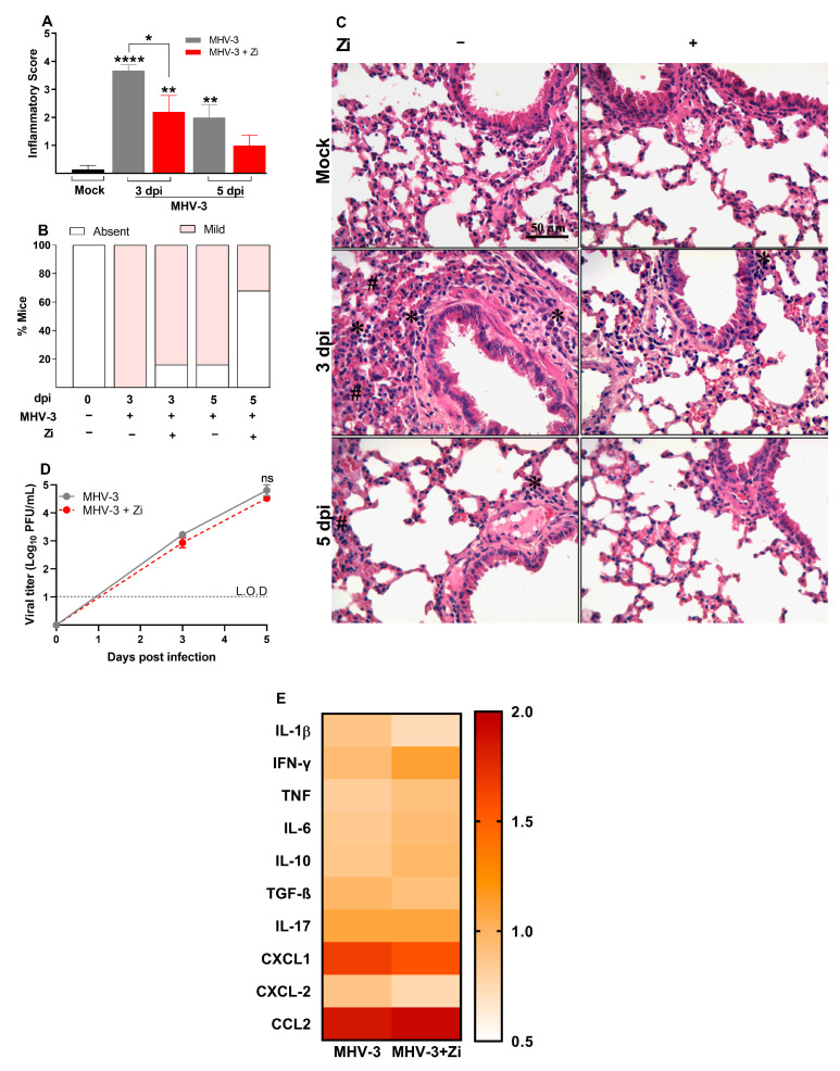
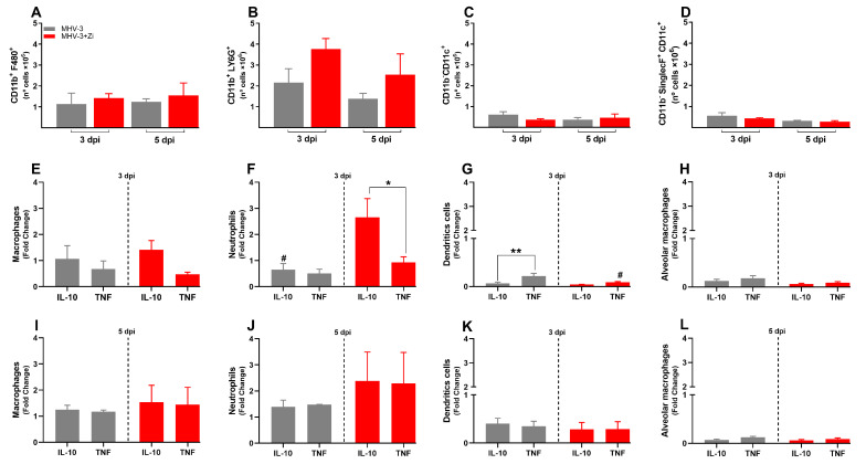
Exacerbated inflammatory responses are a hallmark of severe coronavirus disease 2019 (COVID-19). Zileuton (Zi) is a selective inhibitor of 5-lipoxygenase, an enzyme involved in the production of several inflammatory/pro-resolving lipid mediators. Herein, we investigated the effect of Zi treatment in a severe acute respiratory syndrome (SARS) model. Mouse hepatitis virus (MHV)3-infected mice treated with Zi significantly improved the clinical score, weight loss, cardiopulmonary function, and survival rates compared with infected untreated animals. The protection observed in Zi-treated mice was associated with a lower inflammatory score, reduced dendritic cell-producing tumor necrosis factor (TNF), and increased neutrophil-producing interleukin (IL)-10 in the lungs three days after infection (dpi). At 5 dpi, the lungs of treated mice showed an increase in Th2-, Treg CD4+-, and Treg CD8+-producing IL-10 and reduced Th1 infiltrating cells. Furthermore, similar results were found upon Zi treatment after SARS-CoV-2 infection in transgenic mice expressing the human angiotensin I-converting enzyme 2 (ACE2) receptor driven by the cytokeratin-18 (K18) gene promoter (K18-hACE2), significantly improving the clinical score, weight loss, and lung inflammatory score compared with untreated animals. Our data suggest that Zi protects against developing severe lung disease during SARS induced by betacoronavirus without affecting the host's capacity to deal with infection.

Keywords: COVID-19; MHV; SARS-CoV-2; viral infection; zileuton.


Conflict of interest statement

The authors declare no conflict of interest.

Figures

Figure 1
Figure 1
Kaplan–Meier survival curve of control (Mock), untreated MHV-3-infected, and zileuton (Zi) treated MHV-3-infected mice (Zi: 1.5; 3.0, 15, and 30 mg/mL) (A). Body weight change after infection was assessed by bidirectional repeated measures analysis of variance (ANOVA) and Sidak’s multiple comparison test. Data are presented as mean ± standard error of the mean (SEM) (B). Clinical scoring of control, infected, and Zi-treated infected animals was performed daily until the end of the experiment (C). Viral load was determined in plasma collected from mice infected with MHV-3 using the plaque assay and/or infected and treated with Zi (30 mg/mL). Results are presented as log 10 PFU/μL of plasma. Differences between the groups were assessed using the post hoc Kruskal–Wallis and Dunn test (D). Differential blood count highlighting leukocytes (E), lymphocytes (F), platelets (G), and granulocytes (H), observed on 3 and 5 dpi. Differences between the control, infected, and/or infected-treated groups were assessed by one-way ANOVA and Dunnett’s multiple comparison test. Data are presented as mean ± SEM. LOD, limit of detection. ns, not significant. * p < 0.05; ** p < 0.01; *** p < 0.001; **** p < 0.0001. Abbreviations: MHV-3, mouse hepatitis virus 3; PFU, plaque-forming units.
Figure 2
Figure 2
Histopathological evaluation regarding the general inflammatory score. Comparisons between the Mock and infected groups were performed using the post hoc Kruskal–Wallis and Dunn tests (A). Percentages of mice according to the degree of inflammatory cell infiltration (B). Hematoxylin and eosin (H&E) staining of lung sections showing signs of inflammatory lesion in infected mice (C). * foci inflammatory, # hyperplasia, and H hemorrhage. Viral load was determined in lung extracts of mice infected with MHV-3 and/or infected and treated with zileuton (Zi) using the plaque assay. Results are presented as log 10 PFU/g of tissue (D). Heat map of cytokines and chemokines concentration levels (fold change) in lung homogenates (E). Differences between the groups were assessed using the Kruskal–Wallis and Dunn post hoc tests. LOD, limit of detection. ns, not significant. * p < 0.05; ** p < 0.01; **** p < 0.0001.
Figure 3
Figure 3
Analysis of compliance of the respiratory system (A) in control mice, mice infected with MHV-3 and/or infected and treated with zileuton (Zi). Compliance was calculated from the steepest point of the deflation limb of the pressure-volume (PV) curve. Representative PV curves of animals from each experimental group (mock, MHV-3, and MHV-3+Zi) (B). Percentage of cardiac changes in control mice and mice infected with MHV-3 and/or infected and treated with Zi (C). Computerized electrocardiogram tracings of control mice and mice infected with MHV-3 and/or infected and treated with Zi. Demonstration of the second frontal plane deviation (DII) records, with a speed of 50 mm/s and amplitude 2 N (D). ** p < 0.01.
Figure 4
Figure 4
Flow cytometry analyses showing the total cell numbers in the lung at 3 and 5 dpi among groups of macrophages (CD11b+ F480+) (A), neutrophils (CD11b+ LY6G+) (B), dendritic (CD11b-CD11c+) (C), and macrophages alveolar (CD11b+SinglecF+CD11c+) (D). Macrophages (E,I), neutrophils (F,J), dendritic cells (G,K), and alveolar macrophages (H,L)-producing IL-10 and TNF at 3 and 5 dpi. * p < 0.05; ** p < 0.01; (* representation among the same groups); (# representation between different groups). Abbreviations: IL, interleukin; TNF, tumor necrosis factor.
Figure 5
Figure 5
Flow cytometric analyses showing the total cell numbers in the lung at 3 and 5 dpi between the CD3+CD4+ (A) and CD3+CD8+ (B) groups. CD4+ (C,E) and CD8+ (D,F) cells-producing IFN-γ, IL-17, IL-10, and FOXP3+ IL-10+ (Treg) at 3 and 5 dpi. * p < 0.05; ** p < 0.01; *** p < 0.001. Abbreviations: FOXP3, forkhead box P3; IFN-γ, interferon-gamma; IL, interleukin.
Figure 6
Figure 6
Flow cytometric analyses showing the total cell numbers in the spleen at 3 and 5 dpi among macrophages (CD11b+ F480+) (A), neutrophils (CD11b+ LY6G+) (B), and dendritic (CD11b-CD11c+) (C) groups. Macrophages (D,G), neutrophils (E,H), and dendritic cell-producing (F,I) IL-10 and TNF at 3 and 5 dpi. * p < 0.05; ** p < 0.01 (* representation among the same groups); (# representation between different groups). Abbreviations: IL, interleukin; TNF, tumor necrosis factor.
Figure 7
Figure 7
Flow cytometric analyses showing the total cell numbers in the spleen at 3 and 5 dpi between the CD3+CD4+ (A) and CD3+CD8+ (B) groups. CD4+ (C,E) and CD8+ (D,F) cells-producing IFN-γ, IL-17, IL-10, and Treg at 3 and 5 dpi. * p < 0.05; ** p < 0.01; *** p < 0.001; **** p < 0.0001. Abbreviations: IFN-γ, interferon-gamma; IL, interleukin.
Figure 8
Figure 8
Body weight change after SARS-CoV-2 infection was evaluated using bidirectional repeated measures analysis of variance (ANOVA) and Sidak’s multiple comparison test. Data are presented as mean ± standard error of the mean (SEM) (A). Clinical scoring of control animals, mice infected with SARS-CoV-2, and infected and treated with zileuton (Zi), was performed daily until the end of the experiment (B). Hematoxylin and eosin (H&E) staining of lung sections showing signs of the inflammatory lesions in infected mice (C); * foci inflammatory, # hyperplasia and H hemorrhage. Percentages of mice according to the degree of inflammatory cell infiltration (D). Histopathological evaluation regarding the general inflammatory score. Comparisons between the sham and infection groups were performed using the Kruskal–Wallis and Dunn post hoc tests (E). * p < 0.05; ** p < 0.01; **** p < 0.0001. (* representation among the same groups); (# representation between different groups).

References

    1. Wu C., Chen X., Cai Y., Xia J., Zhou X., Xu S., Huang H., Zhang L., Zhou X., Du C., et al. Risk Factors Associated with Acute Respiratory Distress Syndrome and Death in Patients with Coronavirus Disease 2019 Pneumonia in Wuhan, China. JAMA Intern. Med. 2020;180:934–943. doi: 10.1001/jamainternmed.2020.0994. - DOI - PMC - PubMed
    1. Mehta P., McAuley D.F., Brown M., Sanchez E., Tattersall R.S., Manson J.J. COVID-19: Consider cytokine storm syndromes and immunosuppression. Lancet. 2020;395:1033–1034. doi: 10.1016/S0140-6736(20)30628-0. - DOI - PMC - PubMed
    1. Huang C., Wang Y., Li X., Ren L., Zhao J., Hu Y., Zhang L., Fan G., Xu J., Gu X., et al. Clinical features of patients infected with 2019 novel coronavirus in Wuhan, China. Lancet. 2020;395:497–506. doi: 10.1016/S0140-6736(20)30183-5. - DOI - PMC - PubMed
    1. Arish M., Qian W., Narasimhan H., Sun J. COVID-19 immunopathology: From acute diseases to chronic sequelae. J. Med. Virol. 2023;95:e28122. doi: 10.1002/jmv.28122. - DOI - PMC - PubMed
    1. Mohammed A., Kalle A.M., Reddanna P. Managing SARS-CoV2 Infections through Resolution of Inflammation by Eicosanoids: A Review. J. Inflamm. Res. 2022;15:4349–4358. doi: 10.2147/JIR.S355568. - DOI - PMC - PubMed

Publication types