Review
. 2023 Oct 16;11(10):1600.
doi: 10.3390/vaccines11101600.

In Vitro Transcribed RNA-Based Platform Vaccines: Past, Present, and Future

Affiliations
Review

In Vitro Transcribed RNA-Based Platform Vaccines: Past, Present, and Future

Alexey D Perenkov et al. Vaccines (Basel). .

Abstract

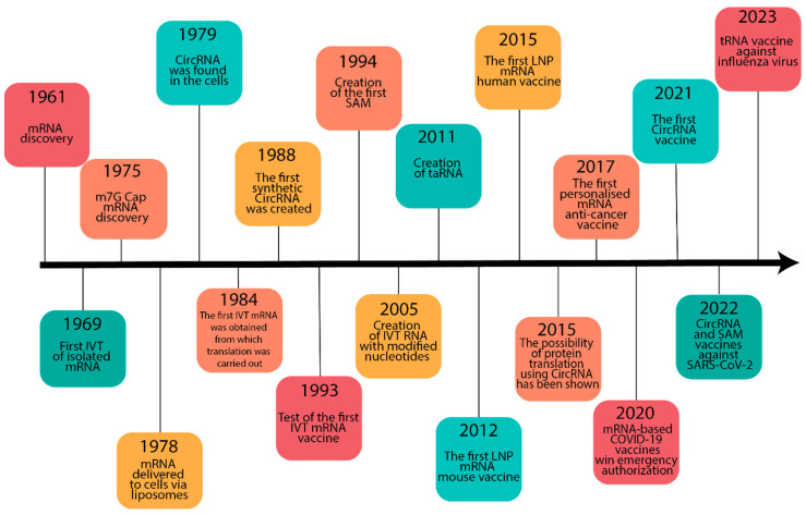
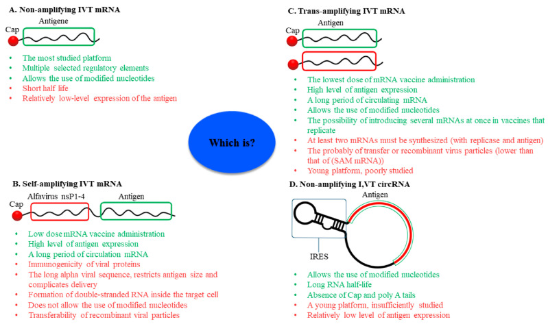
mRNA was discovered in 1961, but it was not used as a vaccine until after three decades. Recently, the development of mRNA vaccine technology gained great impetus from the pursuit of vaccines against COVID-19. To improve the properties of RNA vaccines, and primarily their circulation time, self-amplifying mRNA and trans-amplifying mRNA were developed. A separate branch of mRNA technology is circular RNA vaccines, which were developed with the discovery of the possibility of translation on their protein matrix. Circular RNA has several advantages over mRNA vaccines and is considered a fairly promising platform, as is trans-amplifying mRNA. This review presents an overview of the mRNA platform and a critical discussion of the more modern self-amplifying mRNA, trans-amplifying mRNA, and circular RNA platforms created on its basis. Finally, the main features, advantages, and disadvantages of each of the presented mRNA platforms are discussed. This discussion will facilitate the decision-making process in selecting the most appropriate platform for creating RNA vaccines against cancer or viral diseases.

Keywords: circular RNA vaccine; immunogenicity; mRNA vaccine; self-amplifying mRNA vaccine; trans-amplifying mRNA vaccine; vaccine.


Conflict of interest statement

The authors declare no conflict of interest.

Figures

Figure 1
Figure 1
Timeline of some basic discoveries in the development of RNA vaccine technology. Abbreviations: mRNA, messenger RNA; IVT, in vitro-transcribed; circRNA, circular RNA; SAM, self-amplifying mRNA; taRNA, trans-amplifying mRNA; LNP, lipid nanoparticles; COVID-19, coronavirus disease 2019.
Figure 2
Figure 2
In vitro generation of RNA molecule by a phage T7 RNA polymerase. The blue box represents bacteriophage T7 RNA polymerase recognition site; the red box represents the sequence of interest.
Figure 3
Figure 3
The typical structure of synthetic mRNA. mRNA consists of cap, 5′-untranslated region (5′-UTR), which can include internal ribosome entry sites (IRES), open reading frame (ORF), 3′-untranslated region (3′-UTR), and 3′ poly(A) tail. The colored box characterizes each element in the mRNA structure.
Figure 4
Figure 4
Scheme of cap-dependent and cap-independent initiation of mRNA translation. Cap-dependent translation initiation is the classical way to start the assembly of the eukaryotic initiator complex. The complex includes methionyl initiator tRNA (Met-tRNAi), eukaryotic initiation factor 1 (eIF1), eIF1A, eIF2, eIF3, eIF4A, eIF4B, eIF4G, eIF4E, eIF5, and poly(A)-binding protein (PABP). Class I–IV IRES initiates mRNA translation in a cap-independent way and is typical for viruses. The initiator complex of mRNA translation containing Class I IRES includes all the factors characteristic of the cap-dependent pathway except eIF4E, which recognizes the cap, but adds IRES trans-activating factors (ITAFs). Class II IRES includes all the initiator factors characteristic of Class I IRES, but the initiation itself occurs directly at the AUG codon. IRES class III, similar to IRES class II, attracts translation initiation factors directly to the AUG codon, but only Met-tRNAi, eIF2, and eIF3 perform initiation. Class IV IRES engages the 40S subunit independently without the eIF and AUG codons, promoting translation initiation by structurally mimicking Met-tRNAi.
Figure 5
Figure 5
Schematic representation of the mRNA mechanism of action in promoting the adaptive immune response. mRNA is first internalized into the antigen-presenting cell (1) and an endosome containing the mRNA is formed (2). mRNA must exit the endosome into the cytoplasm to avoid degradation in the phagolysosome (3). Once in the cytoplasm, the mRNA is translated by ribosomes (4). Part of the synthesized antigen is transported to the surface of the antigen-presenting cell (5), where it can be recognized by B-lymphocytes (6), which end up forming clones of plasma cells synthesizing antibodies against the antigen (7). Another portion of the synthesized antigen can be secreted into the intercellular space (8), where it can be captured (9) and degraded as part of the phagolysosome (10) of another antigen-presenting cell. After degradation, the antigen is presented as part of the MHC class II (11). The third pathway of the synthesized antigen is proteasomal degradation into peptides within the synthesizing cell (12). The peptides are then presented as part of the MHC class I (13).
Figure 6
Figure 6
Vaccine design for non-amplifying, self-amplifying, trans-amplifying, and circular in vitro-transcribed RNA (IVT circRNA). Non-amplifying IVT mRNA is the classic RNA vaccine (A). A self-replicating RNA vaccine was created on the basis of non-amplifying IVT mRNA by encoding viral RNA-dependent RNA polymerase (RdRp) (B). The trans-amplifying IVT mRNA vaccine was the logical extension of self-amplifying IVT mRNA. It includes two types of mRNA: mRNA encoding antigens and mRNA encoding viral replicase (С). Circular IVT RNA has a unique closed structure that allows translation initiation only through internal ribosome entry sites (IRES) (D). Green box: RNA fragment encoding the antigen conferring the vaccine properties. Red box: RNA fragment encoding a viral RNA-dependent RNA polymerase that can amplify the antigen-encoding site of the RNA to enhance the vaccine action of a specific dose. IRES: internal ribosome entry sites.
Figure 7
Figure 7
Enzymatic ligation of RNA using T4 ligases. T4 Dnl1 requires the presence of a double-stranded nucleic acid substrate, so a short single-stranded DNA molecule complementary to the circular RNA being synthesized is added to the reaction. T4 Rnl 1 has an affinity for single-stranded RNA molecules. For T4 Rnl 1 to work, the convergence of the joined 3’- and 5’-ends in the RNA molecule is very important, so DNA not fully complementary to the linear RNA molecule is added to the reaction. This DNA splint brings the RNA ends closer together but a few non-complementary terminal nucleotides leave the ends single-stranded. T4 Rnl 2 requires a double-stranded structure to form a circular RNA molecule, so a short RNA or DNA molecule fully complementary to the ends to be joined is added to the reaction.
Figure 8
Figure 8
Ribozyme methods for circRNA synthesis. The group I intron self-splicing system comprises RNA fragments of exons and introns of the foreign organism flanking the antigen sequence. The group I intron self-splicing system requires guanosine triphosphate (GTP) and Mg2+ in the reaction as co-factors for the formation of circRNA from a linear RNA molecule. The principle of the group II intron self-splicing method resembles the principle of the group I intron self-splicing method, but it can use foreign introns without exons. However, the self-splicing of group II introns is known to form 2’, 5’-phosphodiester bonds at the ligation site.
Figure 9
Figure 9
Advantages and disadvantages of different RNA vaccine platforms. Non-amplifying IVT mRNA platform (A), self-amplifying IVT mRNA (B), trans-amplifying IVT mRNA (C), IVT circRNA (D). Green text represents advantages and red text indicates disadvantages of the RNA platform. IVT: in vitro-transcribed, IRES: internal ribosome entry sites.

Similar articles

Cited by

References

    1. Plotkin S.A. Vaccines: The Fourth Century. Clin. Vaccine Immunol. 2009;16:1709–1719. doi: 10.1128/CVI.00290-09. - DOI - PMC - PubMed
    1. Probst J., Weide B., Scheel B., Pichler B.J., Hoerr I., Rammensee H.G., Pascolo S. Spontaneous cellular uptake of exogenous messenger RNA in vivo is nucleic acid-specific, saturable and ion dependent. Gene Ther. 2007;14:1175–1180. doi: 10.1038/sj.gt.3302964. - DOI - PubMed
    1. Ahammad I., Lira S.S. Designing a novel mRNA vaccine against SARS-CoV-2: An immunoinformatics approach. Int. J. Biol. Macromol. 2020;162:820–837. doi: 10.1016/j.ijbiomac.2020.06.213. - DOI - PMC - PubMed
    1. Li W., Joshi M.D., Singhania S., Ramsey K.H., Murthy A.K. Peptide Vaccine: Progress and Challenges. Vaccines. 2014;2:515–536. doi: 10.3390/vaccines2030515. - DOI - PMC - PubMed
    1. Schmidt S.T., Foged C., Korsholm K.S., Rades T., Christensen D. Liposome-Based Adjuvants for Subunit Vaccines: Formulation Strategies for Subunit Antigens and Immunostimulators. Pharmaceutics. 2016;8:7. doi: 10.3390/pharmaceutics8010007. - DOI - PMC - PubMed

LinkOut - more resources