. 2023 Dec 11;51(22):12207-12223.
doi: 10.1093/nar/gkad940.

Human HELQ regulates DNA end resection at DNA double-strand breaks and stalled replication forks

Affiliations

Human HELQ regulates DNA end resection at DNA double-strand breaks and stalled replication forks

Yuqin Zhao et al. Nucleic Acids Res. .

Abstract

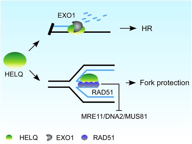
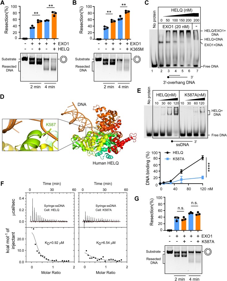
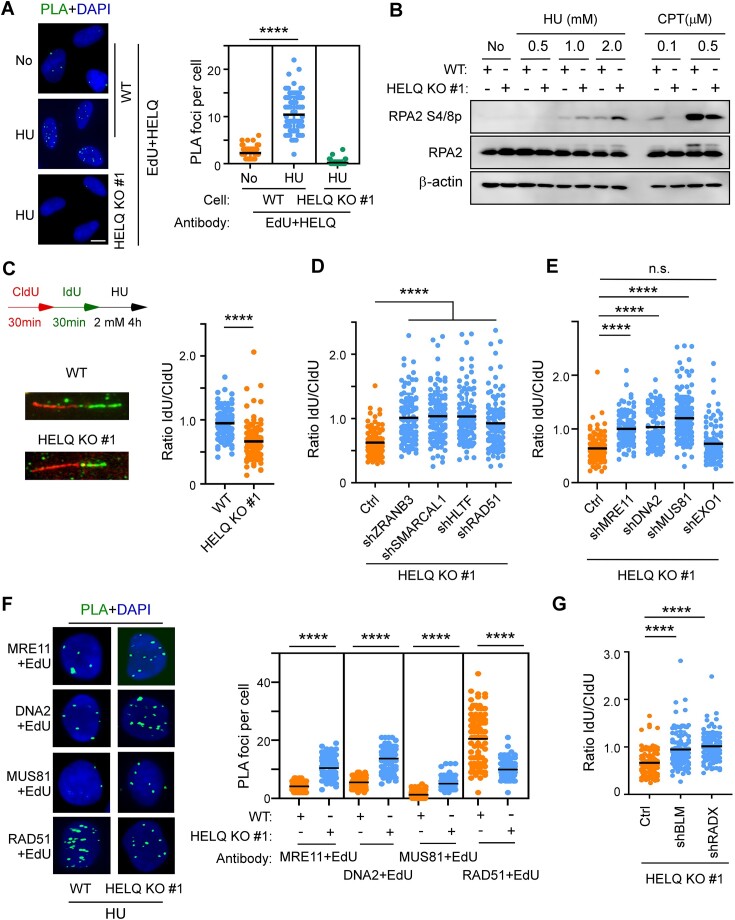
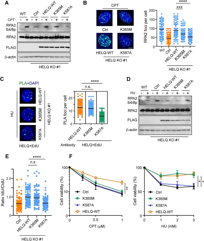
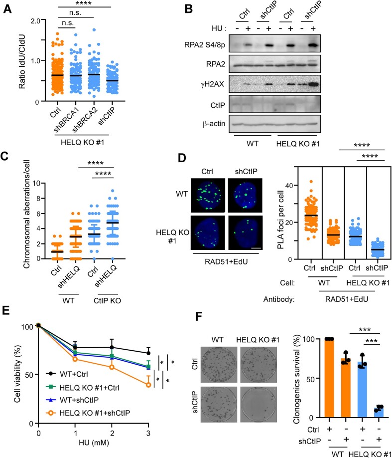
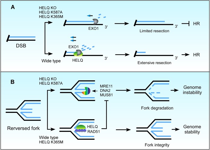
Following a DNA double strand break (DSB), several nucleases and helicases coordinate to generate single-stranded DNA (ssDNA) with 3' free ends, facilitating precise DNA repair by homologous recombination (HR). The same nucleases can act on stalled replication forks, promoting nascent DNA degradation and fork instability. Interestingly, some HR factors, such as CtIP and BRCA1, have opposite regulatory effects on the two processes, promoting end resection at DSB but inhibiting the degradation of nascent DNA on stalled forks. However, the reason why nuclease actions are regulated by different mechanisms in two DNA metabolism is poorly understood. We show that human HELQ acts as a DNA end resection regulator, with opposing activities on DNA end resection at DSBs and on stalled forks as seen for other regulators. Mechanistically, HELQ helicase activity is required for EXO1-mediated DSB end resection, while ssDNA-binding capacity of HELQ is required for its recruitment to stalled forks, facilitating fork protection and preventing chromosome aberrations caused by replication stress. Here, HELQ synergizes with CtIP but not BRCA1 or BRCA2 to protect stalled forks. These findings reveal an unanticipated role of HELQ in regulating DNA end resection at DSB and stalled forks, which is important for maintaining genome stability.


Figures

Graphical Abstract
Graphical Abstract
Figure 1.
Figure 1.
HELQ is recruited to DSBs at the early stages of DSB repair. (A) Time-lapse imaging of EGFP-HELQ and EGFP-Ku70 in U2OS cells before and after microirradiation. The red line marks the damage region. (B) Time-lapse imaging of EGFP-HELQ in U2OS cells expressing vector control (Ctrl) or indicated shRNAs before and after microirradiation. C-E. EGFP-MRE11 (C), EGFP- NBS1 (D) and EGFP-CtIP (E) recruitment was monitored in U2OS (WT) and HELQ KO #1 cells. (F) Top, schematic of ChIP assay in ER-AsiSI U2OS cells. Bottom, SFB-HELQ enrichment at DSBs induced by AsiSI enzyme. ER-AsiSI U2OS cells expressing vector control (Ctrl) or the indicated shRNAs were treated with or without 4-OHT (300 nM, 4 h) to induce DSBs before a ChIP assay was performed using an anti-FLAG antibody. In panel A to E, scale bar = 5 μm. In panel F, the data are derived from three independent experiments and represent the means ± SD. *P < 0.05; n.s.: not significant. Student' s t-test was used.
Figure 2.
Figure 2.
HELQ is required for DSB end resection. (A, B) RPA2 phosphorylation was detected by western blotting after the indicated cells were treated with 2 μM camptothecin (CPT, A) or 10 μM etoposide (ETO, B) for the indicated times. (C) The cells were incubated with BrdU for 24 h, then treated with 2 μM CPT for the indicated times, fixed and then stained with BrdU and γH2AX antibodies under native conditions. Representative images (left) and the quantification of the average number of BrdU foci per γH2AX positive cell (right) are shown. scale bar = 10 μm. (D) Indicated cells were treated with CPT (2 μM, 1 h) followed by RPA2 foci formation analysis by immunostaining with the indicated antibodies. Representative images (top) and the quantification of the average number of RPA2 foci per cell (bottom) are shown. Scale bar = 5 μm. (E) Quantification and schematic of qPCR-based end resection assay in HELQ KO cells (#2 and #3). The data represent the means ± SD of three independent experiments. *P < 0.05, **P < 0.01, ****P < 0.0001. Mann–Whitney test was used in C and D; Student' s t-test was used in E.
Figure 3.
Figure 3.
HELQ and EXO1 act on the same pathway to regulate DSB end resection. A-E. U2OS cells (WT) and HELQ KO #1 cells were infected with lentiviruses encoding the indicated shRNAs or a control vector (Ctrl). RPA2 phosphorylation analysis (A–D) and RPA2 foci formation analysis (E) were performed after CPT (2 μM, 1 h) treatment. Scale bar = 5 μm (E). (F) The HELQ-EXO1 interaction was analyzed by PLA in U2OS cells treated with or without ETO (10 μM, 1 h). Left, representative images. Right, quantification of the average number of PLA foci per nucleus. Scale bar = 10 μm. (G) Time-lapse imaging of EGFP-EXO1 in U2OS (WT) and HELQ KO #1 cells before and after microirradiation. The red line marks the damage region. Scale bar = 5 μm. In panel E and F, the data represent the means ± SD of three independent experiments. ****P < 0.0001, n.s.: not significant. Mann–Whitney test was used.
Figure 4.
Figure 4.
HELQ promotes the nuclease activity of EXO1 in vitro. A-B. Nicked plasmid (7 nM) was incubated with purified wild type HELQ (A) or a helicase-dead HELQ mutant (K365M, (B)) in the presence or absence of EXO1 (15 nM) for the indicated times and the products were resolved on a 0.8% agarose gel. (C) 3′-overhang DNA substrate was incubated with the indicated concentration of HELQ and analyzed in 6% native polyacrylamide gel in the presence or absence of EXO1 (20 nM). Protein-DNA complexes are indicated. (D) A structural model of human HELQ-DNA, constructed by superimposing the structure of Archaeoglobus fulgidus HELQ-DNA with human HELQ using AlphaFold2. The enlarged area shows the interaction between human HELQ K587 and ssDNA. (E) Electrophoretic mobility shift assays such as (C) using HELQ variants and ssDNA substrate. The quantification of DNA binding capacity is shown in the bottom. (F) ITC titration and fitting curves of HELQ variants with ssDNA. (G) The effect of HELQ K587A on EXO1 nuclease as described in (A) and (B). In panels A, B, E and G, error bars represent means ± SD of three independent experiments. **P < 0.01, ****P < 0.0001, n.s.: not significant. Student's t-test.
Figure 5.
Figure 5.
HELQ prevents excessive resection of nascent DNA on the stalled forks. (A) Cells were labelled with EdU for 10 min prior to the addition of 2 mM HU for 4 h. The HELQ-nascent DNA (EdU) interaction was analyzed by PLA. Quantification of the average number of PLA foci per nucleus is shown on the right. Scale bar = 10 μm. (B) U2OS (WT) and HELQ KO #1 cells were treated with the indicated drugs (HU and CPT for 4 h) followed by RPA2 phosphorylation analysis by western blotting. (C) Fork degradation assays were performed in U2OS (WT) and HELQ KO #1 cells. Left, schematic of the assays and representative fiber images for the indicated samples. Right, the scatterplot of IdU-/CldU- tract length ratios for individual replication fork was shown. (D and E) The same fork degradation assays as in C were performed in HELQ KO #1 cells expressing vector control (Ctrl) or indicated shRNAs. The scatterplot of IdU-/CldU- tract length ratios for individual replication forks is shown. (F) Interactions between the indicated proteins and nascent DNA (EdU) were detected as described in A. Representative PLA images and quantification of the average number of PLA foci per nucleus detected by the indicated antibodies are shown. Scale bar = 5 μm. (G) Fork degradation assays as in C were performed in HELQ KO #1 cells expressing vector control (Ctrl) or the indicated shRNAs. A scatterplot of IdU-/CldU- tract length ratios for individual replication forks is shown. In panels A, C, D, E, F and G, the data are representative of at least three independent experiments. **** P < 0.0001, n.s.: not significant. Mann–Whitney test.
Figure 6.
Figure 6.
The ssDNA binding activity of HELQ is important for DSB end resection and fork protection. A-B. HELQ KO #1 cells expressing vector control or the indicated HELQ variants were subjected to CPT (2 μM, 2 h) induced RPA2 phosphorylation (A) and RPA2 foci formation (B) assays. Scale bar = 5 μm (B). (C–E) HELQ-EdU PLA assay, RPA2 phosphorylation (D) and fork degradation assay (E) (C-E: 2 mM HU for 4 h,) were performed in HELQ KO #1 cells expressing the indicated HELQ variants. Scale bar = 5 μm (C). F. HELQ KO #1 cells expressing vector control or the indicated HELQ variants were treated with the indicated concentrations of CPT (left) or HU (right) for 48 h, then a cell viability assay was performed. In panels B, C, E and F, data represent the means ± SD from three independent experiments. *P < 0.05, **P < 0.01, ****P < 0.0001. n.s.: not significant. Mann–Whitney test was used in B, C and D; Student's t-test was used in F.
Figure 7.
Figure 7.
HELQ functions in parallel to CtIP to preserve fork integrity. (A) Fork degradation assays were performed in HELQ KO #1 cells expressing the indicated shRNAs. (B) HU (2 mM, 4 h) induced RPA2 phosphorylation assays were performed in wild type (WT) and HELQ KO #1 cells expressing shCtIP or vector (Ctrl). (C) HCT116 cells (WT) and HCT116 CtIP KO cells (CtIP KO) expressing shHELQ or vector (Ctrl) were treated with 2 mM HU for 4 h and subjected to metaphase spread assay. The chromosomal aberrations per chromosome spread are plotted. (D) The HU (2 mM, 4 h) induced RAD51-nascent DNA (EdU) interaction was analyzed by PLA in U2OS cells (WT) and HELQ KO #1 cells expressing shCtIP or vector (Ctrl). Representative images and quantification of the average number of PLA foci per nucleus are shown. Scale bar = 5 μm. (E) WT and HELQ KO #1 cells expressing shCtIP or vector (Ctrl) were treated with the indicated concentration of HU for 48 h, then a cell viability assay was performed. (F) The viability of WT and HELQ KO #1 cells expressing shCtIP or vector (Ctrl) was analyzed by colony formation assay. Representative images (left) and quantifications of the relative survival (right) relative to vector (Ctrl) infected wild type U2OS cells (WT), which was arbitrarily set to 100%, are shown. In panels A, C, D, E and F, the data represent the means ± SD from at least three independent experiments. *P < 0.05, ***P < 0.001, ****P < 0.0001, n.s.: not significant. Mann–Whitney test was used in A, C and D; Student's t-test was used in E and F.
Figure 8.
Figure 8.
A model illustrating the roles of HELQ in DSB end resection and fork protection. (A) HELQ promotes EXO1-mediated DSB end resection. In the presence of HELQ, EXO1 drives extensive end resection, which is necessary for HR. Without HELQ, EXO1 activity is not fully stimulated, and the ssDNA generated by end resection is insufficient to initiate HR. Both HELQ helicase activity and ssDNA binding ability are needed to promote EXO1 activity in cells. (B) HELQ prevents degradation of nascent DNA on reversed replication forks. HELQ stabilizes RAD51 at reversed forks and limits fork resection by nucleases. Loss of HELQ or its ssDNA binding capacity leads to fork degradation and genomic instability.

References

    1. Aguilera A., Gomez-Gonzalez B.. Genome instability: a mechanistic view of its causes and consequences. Nat. Rev. Genet. 2008; 9:204–217. - PubMed
    1. White R.R., Vijg J.. Do DNA double-strand breaks drive aging?. Mol. Cell. 2016; 63:729–738. - PMC - PubMed
    1. Gennery A.R., Cant A.J., Jeggo P.A.. Immunodeficiency associated with DNA repair defects. Clin. Exp. Immunol. 2000; 121:1–7. - PMC - PubMed
    1. McKinnon P.J. DNA repair deficiency and neurological disease. Nat. Rev. Neurosci. 2009; 10:100–112. - PMC - PubMed
    1. Moynahan M.E., Jasin M.. Mitotic homologous recombination maintains genomic stability and suppresses tumorigenesis. Nat. Rev. Mol. Cell Bio. 2010; 11:196–207. - PMC - PubMed

Publication types