. 2023 Nov 27;51(21):11941-11951.
doi: 10.1093/nar/gkad951.

Structural and functional investigation of GajB protein in Gabija anti-phage defense

Affiliations

Structural and functional investigation of GajB protein in Gabija anti-phage defense

Hyejin Oh et al. Nucleic Acids Res. .

Abstract

Bacteriophages (phages) are viruses that infect bacteria and archaea. To fend off invading phages, the hosts have evolved a variety of anti-phage defense mechanisms. Gabija is one of the most abundant prokaryotic antiviral systems and consists of two proteins, GajA and GajB. GajA has been characterized experimentally as a sequence-specific DNA endonuclease. Although GajB was previously predicted to be a UvrD-like helicase, its function is unclear. Here, we report the results of structural and functional analyses of GajB. The crystal structure of GajB revealed a UvrD-like domain architecture, including two RecA-like core and two accessory subdomains. However, local structural elements that are important for the helicase function of UvrD are not conserved in GajB. In functional assays, GajB did not unwind or bind various types of DNA substrates. We demonstrated that GajB interacts with GajA to form a heterooctameric Gabija complex, but GajB did not exhibit helicase activity when bound to GajA. These results advance our understanding of the molecular mechanism underlying Gabija anti-phage defense and highlight the role of GajB as a component of a multi-subunit antiviral complex in bacteria.


Figures

Graphical Abstract
Graphical Abstract
Figure 1.
Figure 1.
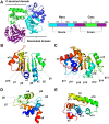
Crystal structure of GajB reveals a four-subdomain architecture. (A) Overall structure of GajB. Ncore, Nacc, Ccore and Cacc subdomains of GajB are in cyan, blue, purple and green, respectively. Schematic linear representation of GajB subdomain organization is also shown with residue numbers indicating the subdomain boundaries. (B–E) Structures of the Ncore (B), Ccore (C), Nacc (D) and Cacc (E) subdomains of GajB. Each subdomain is shown in rainbow format from the N terminus (blue) to the C terminus (red). Secondary structural elements are also indicated.
Figure 2.
Figure 2.
Structural similarity between GajB and UvrD. (A) Side-by-side comparison of GajB and UvrD (PDB ID: 3LFU) structures. GajB and UvrD share a four-subdomain architecture. Subdomains of GajB are colored as in Figure 1. The 1A, 1B, 2A and 2B subdomains of UvrD are shown in red, orange, yellow and brown, respectively. (B–D) Structural alignments of subdomains between GajB and UvrD. The Ncore (B) and Ccore (C) subdomains of GajB are superposed well with the 1A and 2A subdomains of UvrD, respectively. Structural alignment of the Nacc (D) subdomain in GajB with the 1B subdomain of UvrD reveals only a partial similarity. Structural superposition is not shown for the Cacc subdomain of GajB and 2B subdomain of UvrD because there was no significant match between them.
Figure 3.
Figure 3.
GajB lacks functionally important structural elements of UvrD. The gating helix (magenta), GIG motif (green) and separation pin (yellow) are structural elements that are important for helicase activity of UvrD. These three elements are highlighted in the structure of UvrD (PDB ID: 3LFU; grey). In close-up views, the corresponding parts in the GajB structure (blue) are superposed for comparison.
Figure 4.
Figure 4.
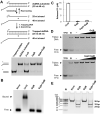
Activity assays of Gabija proteins. (A) Helicase activity assay of GajB and GajA:GajB complex. 3′-Tailed dsDNA substrates were incubated with proteins and analyzed by PAGE. Asterisks indicate fluorescein labels. (B) DNA binding assay of GajB and GajA:GajB complex. Agarose-based EMSAs of proteins (10.0 μM each) were conducted with 3′-tailed dsDNA substrate. (C) ATPase activity assay of GajB and GajA:GajB complex. Amounts of released inorganic phosphate from three independent experiments are shown as mean ± standard error of the mean (SEM). (D) DNA translocase activity assays of GajB and GajA:GajB complex. DNA translocation was analyzed by triplex displacement assay, in which fluorescein-labeled triplex forming oligonucleotide (TFO) was hybridized to linear dsDNA, and translocation was measured after the addition of ATP and proteins (0.5, 2.5 and 10.0 μM each). Samples were analyzed by PAGE. ‘B’ represents boiled sample. (E) Nuclease activity assay of GajA and GajA:GajB complex. Proteins were incubated with DNA substrate containing GajA recognition sequence. Reaction products were analyzed by PAGE and visualized by ethidium bromide. The sequences of oligonucleotides used in the assays are listed in Table S1. Uncropped gel images are shown in Figure S8.
Figure 5.
Figure 5.
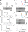
GajB interacts with GajA to form a 4:4 Gabija complex. (A) Co-elution of co-expressed GajA and GajB in SEC. When GajA and GajB were expressed together in E. coli, they co-purified in SEC. (B) Formation of Gabija complex from individually purified GajA and GajB proteins in SEC. When incubated together prior to injection, individually purified GajA and GajB proteins migrated together (purple) during SEC with a smaller retention volume than those for GajA (red) and GajB (blue). (C, D) SEC-MALS analyses of GajA and GajB. Oligomeric states of GajA (C) and GajB (D) were investigated by SEC-MALS. The average molar masses corresponding the SEC peaks are consistent with tetrameric GajA and monomeric GajB. Theoretical molecular weights of the GajA tetramer and the GajB monomer used in these experiments are 272.2 and 57.3 kDa, respectively. (E) SEC analysis of the stoichiometry of Gabija complex. GajA was pre-incubated with increasing amounts of GajB, and the mixtures were analyzed by SEC. Until the molar ratio of GajA to GajB reached 4:4 (cyan and orange lines), the retention volume decreased and the peak height of the complex increased. Addition of extra GajB (black dashed line) did not change the peak of the complex, but resulted in emergence of an unbound GajB peak. (F) SEC-MALS analysis of Gabija complex. GajA was pre-incubated with an excess amount of GajB, and the mixture was analyzed by SEC-MALS. The molar ratio of GajA to GajB was 4:8. The average molar masses corresponding the two SEC peaks were consistent with the 4:4 GajA:GajB complex and monomeric GajB. The theoretical molecular weight of the 4:4 GajA:GajB complex used in this experiment is 501.3 kDa (G) ITC trace for GajA binding to GajB. The isotherm is representative of triplicate measurements. The dissociation constant (Kd) and binding ratio (N) are presented as mean ± SEM. Data for the three independent ITC experiments are shown in Figure S6. Uncropped gel images are shown in Figure S8.

References

    1. Ofir G., Sorek R.. Contemporary phage biology: from classic models to new insights. Cell. 2018; 172:1260–1270. - PubMed
    1. Fuhrman J.A. Marine viruses and their biogeochemical and ecological effects. Nature. 1999; 399:541–548. - PubMed
    1. Weinbauer M.G. Ecology of prokaryotic viruses. FEMS Microbiol. Rev. 2004; 28:127–181. - PubMed
    1. Stern A., Sorek R.. The phage-host arms race: shaping the evolution of microbes. Bioessays. 2011; 33:43–51. - PMC - PubMed
    1. Hampton H.G., Watson B.N.J., Fineran P.C.. The arms race between bacteria and their phage foes. Nature. 2020; 577:327–336. - PubMed

Publication types