. 2023 Dec;22(12):2861-2875.
doi: 10.1007/s43630-023-00493-3. Epub 2023 Oct 28.

Growth, morphology, and formation of cinnabarin in Pycnoporus cinnabarinus in relation to different irradiation spectra

Affiliations

Growth, morphology, and formation of cinnabarin in Pycnoporus cinnabarinus in relation to different irradiation spectra

Christoph W Schinagl et al. Photochem Photobiol Sci. 2023 Dec.

Abstract

Background: The demand for natural pigments in general, and for fungi-derived pigments in particular, is constantly rising. Wood-decomposing fungi represent a promising source for natural pigments and they are usually easy to cultivate in pure culture. One of them, i.e., Pycnoporus cinnabarinus, offers a highly interesting spectrum of bioactivity, partly due to the formation of the orange-red pigment cinnabarin. However, apart from a few studies addressing its diverse potential biotechnological applications, there is still a large gap of knowledge concerning the influence of light on the formation of cinnabarin. The aim of this work was to investigate the effect of different irradiations on the cinnabarin content, the growth, and the morphology of three different P. cinnabarinus strains. We used highly standardized irradiation conditions and cultivation techniques in combination with newly developed methods for the extraction and direct quantification of cinnabarin.

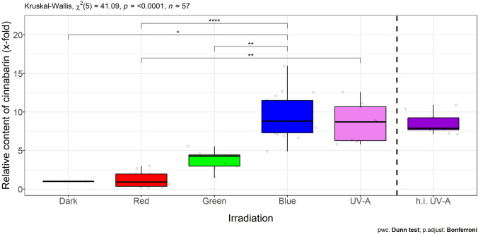
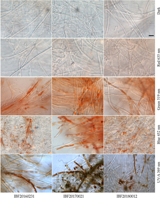
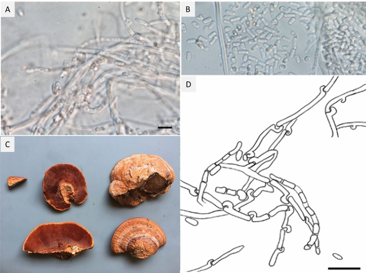
Results: Red, green, blue, and UV-A irradiation (mean irradiance Ee = 1.5 ± 0.18 W m-2) had considerable effects on the growth and colony appearance of all three P. cinnabarinus strains tested. The cinnabarin content determined was, thus, dependent on the irradiation wavelength applied, allowing strain-specific thresholds to be defined. Irradiation with wavelengths below this strain-specific threshold corresponded to a lower cinnabarin content, at least at the intensity applied. The orange-red pigment appeared by light microscopy as incrusted extracellular plaques present on the hyphal walls. Highly efficient vegetative propagation occurred by arthroconidia, and we observed the tendency that this asexual reproduction was (i) most frequent in the dark but (ii) never occurred under UV-A exposure.

Conclusion: This study highlights a differential photo-dependence of growth, morphology, and cinnabarin formation in P. cinnabarinus. This confirms that it is advisable to consider the wavelength of the light used in future biotechnological productions of natural pigments.

Keywords: Arthroconidia; Cinnabarin; LIGHT BOX; Light-induced pigmentation; Natural product; Standardized irradiation conditions; Wood-inhabiting fungi.


Conflict of interest statement

The authors declare no conflict of interest. The funders had no role in the design of the study; in the collection, analyses, or interpretation of data; in the writing of the manuscript; or in the decision to publish the results.

Figures

Fig. 1
Fig. 1
Radial growth (5 days in darkness on MEA) of three P. cinnabarinus strains in dependence of the temperature. Means and standard deviations (n = 3) of the culture diameters of each strain are shown
Fig. 2
Fig. 2
Formation of biomass (mg DW per Petri dish) in cultures of three strains of P. cinnabarinus (IBF20160231, IBF20170021, and IBF20180012) grown on MEA (7 days at 25 °C) in dependence of the following continuous irradiation conditions. Wavelength of peak emission, λpeak, and full width half maximum (FWHM) are indicated in brackets: red (635 ± 18 nm), green (519 ± 38 nm), blue (452 ± 19 nm), and UV-A (369 ± 13 nm). Irradiance for light spectra in the center of the Petri dishes was Ee = 1.5 ± 0.18 W m−2. Dark conditions served as control. Median and standard deviation are depicted; the light gray circles represent individual measurements of cultures
Fig. 3
Fig. 3
Content of cinnabarin (mg g−1 DW, mean sd) in cultures of three P. cinnabarinus strains (IBF20160231, IBF20170021, and IBF20180012) grown on MEA (7 days at 25 °C) in dependence of the following continuous irradiation conditions. Wavelength of peak emission, λpeak, and FWHM are indicated in brackets: red (635 ± 18 nm), green (519 ± 38 nm), blue (452 ± 19 nm), and UV-A (369 ± 13 nm). Average irradiance for light spectra in the center of the Petri dishes was Ee = 1.5 ± 0.18 W m−2
Fig. 4
Fig. 4
Relative content of cinnabarin (x-fold) with respect to content under dark or red conditions, respectively (= onefold) of three P. cinnabarinus strains (IBF20160231, IBF20170021, and IBF20180012) grown on MEA (7 days at 25 °C) in dependence of the following continuous irradiation conditions. Wavelength of peak emission, λpeak, and FWHM are indicated in brackets: red (635 ± 18 nm), green (519 ± 38.1 nm), blue (452 ± 18.7 nm), and UV-A (369 ± 13 nm). Irradiance in the center of the Petri dishes was Ee = 1.5 ± 0.18 W m−2 for all irradiations, except for higher intensity (h.i.) UV-A (369 ± 13 nm) irradiance: Ee = 5.6 ± 1.8 W m−2. Dark conditions served as control for light treatment or base line for the constitutive content of cinnabarin, respectively. Median and standard deviation are depicted, the light gray circles represent individual measurements of cultures
Fig. 5
Fig. 5
Extracellular aggregation of pigments, band-like increased at the cell wall, in dependence of different irradiation conditions (bright field microscopy, 1000-fold magnification). The scale bar equals 10 µm. Irradiance in the center of the Petri dishes was Ee = 1.5 ± 0.18 W m−2 for all irradiations, except for UV-A (369 ± 13 nm): Ee = 5.6 ± 1.8 W m−2
Fig. 6
Fig. 6
Pycnoporus cinnabarinus. A Formation of unpigmented arthroconidia in pure cultures of strain IBF20180012 and B strain IBF20160231. C Fruiting body of Pycnoporus cinnabarinus IBF20160231. D Drawing of arthroconidia formation. A, B, D: scale bar represents 10 µm

Similar articles

Cited by

References

    1. Aly AH, Debbab A, Proksch P. Fungal endophytes: Unique plant inhabitants with great promises. Applied Microbiology and Biotechnology. 2011;90(6):1829–1845. doi: 10.1007/s00253-011-3270-y. - DOI - PubMed
    1. Saleem M, Nazir M. Chapter 9—Bioactive natural products from marine-derived fungi: An update. In: Atta ur R, editor. Studies in natural products chemistry. Elsevier; 2015. pp. 297–361.
    1. Dufosse L, Fouillaud M, Caro Y, Mapari SAS, Sutthiwong N. Filamentous fungi are large-scale producers of pigments and colorants for the food industry. Current Opinion in Biotechnology. 2014;26:56–61. doi: 10.1016/j.copbio.2013.09.007. - DOI - PubMed
    1. Lagashetti AC, Dufossé L, Singh SK, Singh PN. Fungal pigments and their prospects in different industries. Microorganisms. 2019;7(12):604. doi: 10.3390/microorganisms7120604. - DOI - PMC - PubMed
    1. Siewert B. Does the chemistry of fungal pigments demand the existence of photoactivated defense strategies in basidiomycetes? Photochemical and Photobiological Sciences. 2021;20(4):475–488. doi: 10.1007/s43630-021-00034-w. - DOI - PubMed

Supplementary concepts

LinkOut - more resources