Randomized Controlled Trial
. 2024 Mar;51(4):1121-1132.
doi: 10.1007/s00259-023-06467-y. Epub 2023 Oct 28.

Intra-arterial peptide-receptor radionuclide therapy for neuro-endocrine tumour liver metastases: an in-patient randomised controlled trial (LUTIA)

Affiliations
Randomized Controlled Trial

Intra-arterial peptide-receptor radionuclide therapy for neuro-endocrine tumour liver metastases: an in-patient randomised controlled trial (LUTIA)

S C Ebbers et al. Eur J Nucl Med Mol Imaging. 2024 Mar.

Abstract

Purpose: Peptide receptor radionuclide therapy (PRRT) using [177Lu]Lu-DOTATATE has been shown to effectively prolong progression free survival in grade 1-2 gastroenteropancreatic neuroendocrine tumours (GEP-NET), but is less efficacious in patients with extensive liver metastases. The aim was to investigate whether tumour uptake in liver metastases can be enhanced by intra-arterial administration of [177Lu]Lu-DOTATATE into the hepatic artery, in order to improve tumour response without increasing toxicity.

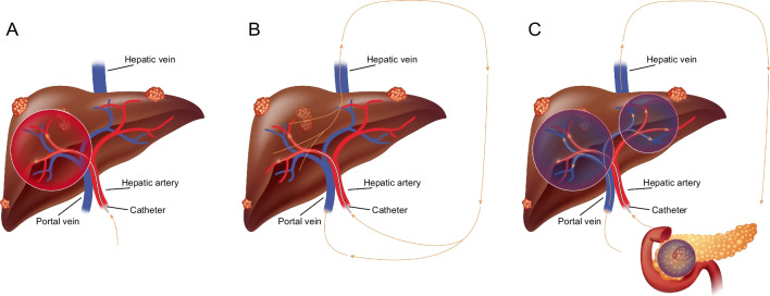
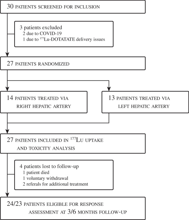
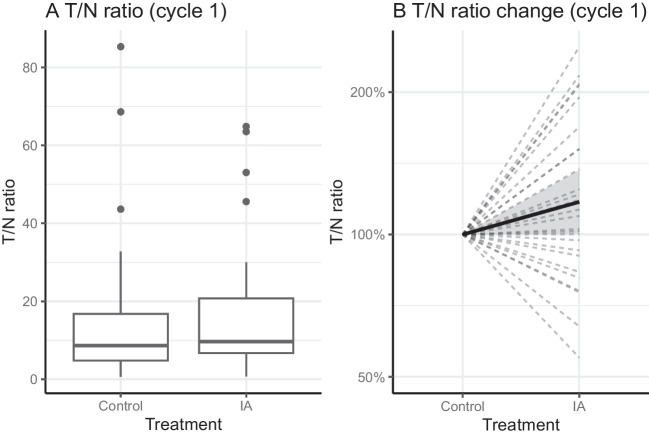
Methods: Twenty-seven patients with grade 1-2 GEP-NET, and bi-lobar liver metastases were randomized to receive intra-arterial PRRT in the left or right liver lobe for four consecutive cycles. The contralateral liver lobe and extrahepatic disease were treated via a "second-pass" effect and the contralateral lobe was used as the control lobe. Up to three metastases (> 3 cm) per liver lobe were identified as target lesions at baseline on contrast-enhanced CT. The primary endpoint was the tumour-to-non-tumour (T/N) uptake ratio on the 24 h post-treatment [177Lu]Lu-SPECT/CT after the first cycle. This was calculated for each target lesion in both lobes using the mean uptake. T/N ratios in both lobes were compared using paired-samples t-test.

Findings: After the first cycle, a non-significant difference in T/N uptake ratio was observed: T/NIA = 17·4 vs. T/Ncontrol = 16·2 (p = 0·299). The mean increase in T/N was 17% (1·17; 95% CI [1·00; 1·37]). Of all patients, 67% (18/27) showed any increase in T/N ratio after the first cycle.

Conclusion: Intra-arterial [177Lu]Lu-DOTATATE is safe, but does not lead to a clinically significant increase in tumour uptake.

Keywords: Efficacy; Intra-arterial; Neuroendocrine tumour; PRRT; Safety.


Conflict of interest statement

The authors declare no competing interests.

Figures

Fig. 1
Fig. 1
A Intra-arterial administration was performed in either the left or the right hepatic artery, thereby exposing half of the liver to high concentration [177Lu]Lu-DOTATATE. B [177Lu]Lu-DOTATATE enters systemic circulation via the hepatic vein. C tumours in the contra-lateral lobe and extrahepatic disease are treated via systemic circulation, both via portal vein and hepatic artery (second-pass effect)
Fig. 2
Fig. 2
Example procedure in a 64 years old male patient with grade I small-intestinal NET, with lymphatic, hepatic, pulmonary, orbital and skeletal metastases. The patient was randomized to intra-arterial PRRT infused from the left hepatic artery. A: baseline CECT depicting extensive bi-lobar disease; B: baseline [68Ga]Ga-DOTATOC PET/CT maximum intensity projection depicting extensive metastatic disease; C: cone-beam CT showing the microcatheter placed in the left hepatic artery, and hypervascular tumours in the left liver lobe; D: post-treatment 177Lu SPECT/CT, showing similar activity distribution compared to the baseline [68Ga]Ga-DOTATOC PET/CT. The resulting T/NIA ratio was 9.4, and the T/Ncontrol ratio was 6.9. Both liver lobes showed stable disease at 3 and 6 months post-treatment follow-up imaging
Fig. 3
Fig. 3
Flowchart of patient inclusion, treatment and analysis. Due to the in-patient randomization, no dedicated flowchart per treatment arm can be shown. One patient died due to disease progression after receiving the first cycle. One patient was referred for additional treatment before finishing four cycles of intra-arterial PRRT, and another patient was referred after the 3-month follow-up imaging. One patient voluntarily withdrew from the study after the first cycle. Twenty-four patients finished all four treatment cycles
Fig. 4
Fig. 4
A Boxplot of the T/N uptake ratios observed in control and intra-arterially treated liver metastases, calculated on the 24 h post-treatment SPECT/CT after the first cycle (primary endpoint). B Uptake in intra-arterially treated liver metastases relative to control tumours. Mean relative change = 17% (p = 0·045, 95% CI [0·00, 0·31]). T/N = Tumour-to-non-tumour; IA = Intra-arterial
Fig. 5
Fig. 5
Response of hepatic metastases after intra-arterial treatment. A: trend of mean diameter of liver metastases with confidence interval from baseline to 6 months post-treatment. B and C: Waterfall plots showing the relative change in diameter of all target lesions in each liver lobe compared to baseline. Intra-arterially treated lobes and control lobes are distributed rather uniformly across the plot, indicating no significant difference in response. Note: each subject is present in each waterfall plot twice, due to intra-patient comparison. IA = Intra-arterial
Fig. 6
Fig. 6
Mean hepatic parameters during follow-up after intra-arterial administration of [177Lu]Lu-DOTATATE show a slight decrease of pre-existent elevated liver enzymes and no indication of additional hepatic toxicity

Similar articles

Cited by

References

    1. Lepage C, Bouvier AM, Faivre J. Endocrine tumours: epidemiology of malignant digestive neuroendocrine tumours. Eur J Endocrinol. 2013;168:R77–R83. doi: 10.1530/EJE-12-0418. - DOI - PubMed
    1. Niederle MB, Hackl M, Kaserer K, Niederle B. Gastroenteropancreatic neuroendocrine tumours: the current incidence and staging based on the WHO and European Neuroendocrine Tumour Society classification: an analysis based on prospectively collected parameters. Endocr Relat Cancer. 2010;17:909–918. doi: 10.1677/ERC-10-0152. - DOI - PubMed
    1. Lesén E, Granfeldt D, Berthon A, Dinet J, Houchard A, Myrenfors P, et al. Treatment patterns and survival among patients with metastatic gastroenteropancreatic neuroendocrine tumours in Sweden-a population-based register-linkage and medical chart review study. J Cancer. 2019;10:6876–6887. doi: 10.7150/jca.32381. - DOI - PMC - PubMed
    1. Sonbol MB, Mazza GL, Mi L, Oliver T, Starr J, Gudmundsdottir H, et al. Survival and incidence patterns of pancreatic neuroendocrine tumors over the last 2 decades: a SEER database analysis. Oncologist. 2022;27:573–578. doi: 10.1093/ONCOLO/OYAC049. - DOI - PMC - PubMed
    1. Frilling A, Modlin IM, Kidd M, Russell C, Breitenstein S, Salem R, et al. Recommendations for management of patients with neuroendocrine liver metastases. Lancet Oncol. 2014;15:e8–21. doi: 10.1016/S1470-2045(13)70362-0. - DOI - PubMed

Publication types

MeSH terms