Review
. 2023 Dec;299(12):105397.
doi: 10.1016/j.jbc.2023.105397. Epub 2023 Oct 28.

ADP-ribose contributions to genome stability and PARP enzyme trapping on sites of DNA damage; paradigm shifts for a coming-of-age modification

Affiliations
Review

ADP-ribose contributions to genome stability and PARP enzyme trapping on sites of DNA damage; paradigm shifts for a coming-of-age modification

Élise Rouleau-Turcotte et al. J Biol Chem. 2023 Dec.

Abstract

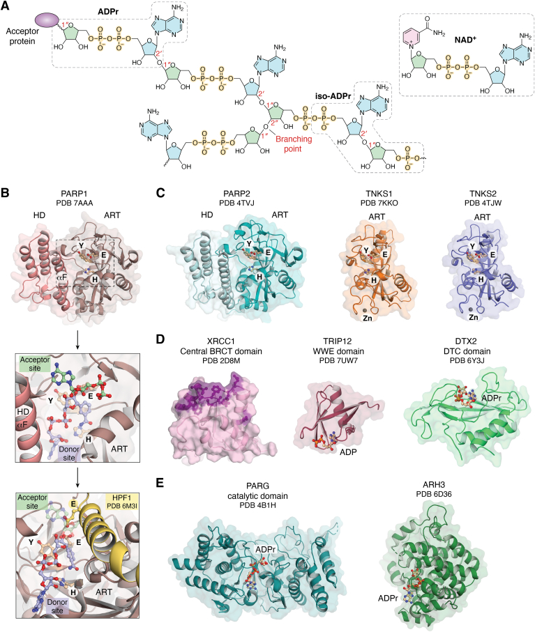
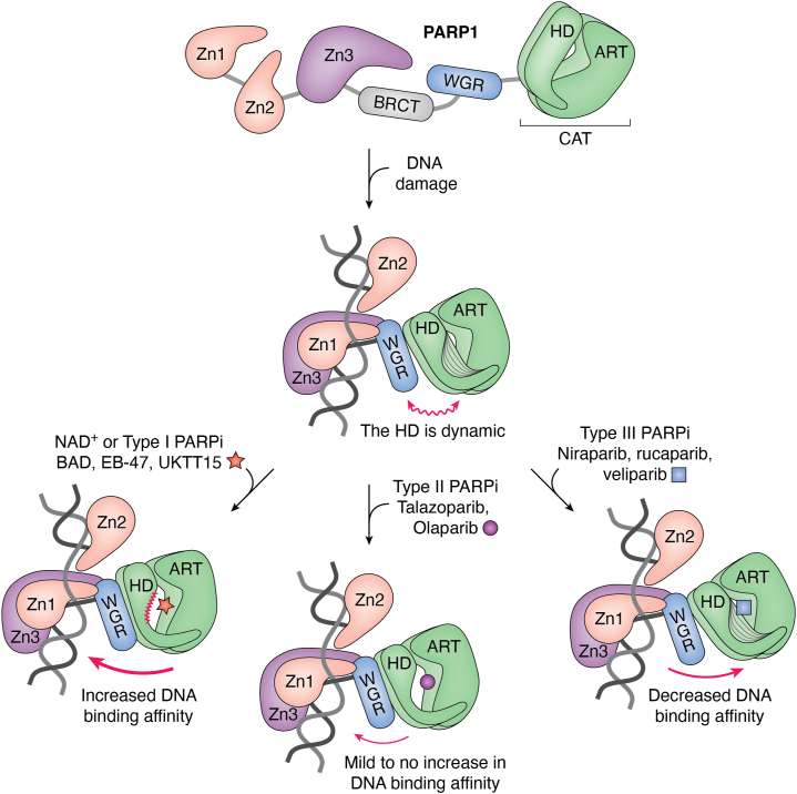
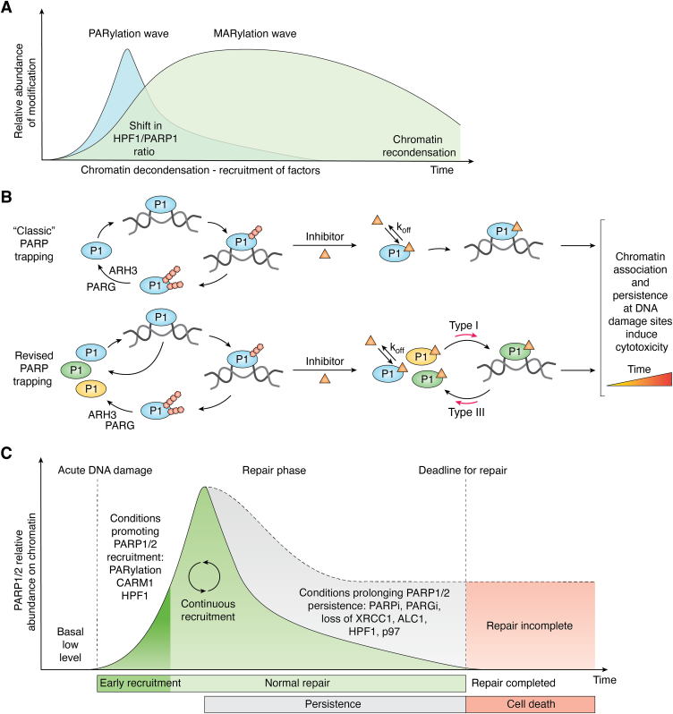
ADP-ribose is a versatile modification that plays a critical role in diverse cellular processes. The addition of this modification is catalyzed by ADP-ribosyltransferases, among which notable poly(ADP-ribose) polymerase (PARP) enzymes are intimately involved in the maintenance of genome integrity. The role of ADP-ribose modifications during DNA damage repair is of significant interest for the proper development of PARP inhibitors targeted toward the treatment of diseases caused by genomic instability. More specifically, inhibitors promoting PARP persistence on DNA lesions, termed PARP "trapping," is considered a desirable characteristic. In this review, we discuss key classes of proteins involved in ADP-ribose signaling (writers, readers, and erasers) with a focus on those involved in the maintenance of genome integrity. An overview of factors that modulate PARP1 and PARP2 persistence at sites of DNA lesions is also discussed. Finally, we clarify aspects of the PARP trapping model in light of recent studies that characterize the kinetics of PARP1 and PARP2 recruitment at sites of lesions. These findings suggest that PARP trapping could be considered as the continuous recruitment of PARP molecules to sites of lesions, rather than the physical stalling of molecules. Recent studies and novel research tools have elevated the level of understanding of ADP-ribosylation, marking a coming-of-age for this interesting modification.

Keywords: ADP-ribose; ADP-ribosyltransferase; ARH3; DNA damage; DNA strand breaks; PARG; PARP enzymes; PARP inhibitors; PARP trapping; allostery; chromatin; poly(ADP-ribose); poly(ADP-ribose) glycohydrolases; protein–nucleic acid interactions.


Conflict of interest statement

Conflict of interest J. M. P. is a cofounder of Hysplex LLC with interests in PARPi development. É. R.-T. declares no conflict of interest with the contents of this article.

Figures

Figure 1
Figure 1
The ADPr/PAR modification and proteins involved in its synthesis, turnover, and signaling.A, schematic representation of a PAR chain composed of (2′-1″) ribose–ribose glycosidic bonds (chain elongation) and occasionally a (2″-1″) ribose–ribose bond (branching point). The modification is attached to target proteins via the anomeric C1″ atom of the first ADPr moiety. The ADPr moiety represents the repeated unit of a PAR chain. Iso-ADPr is highlighted as well as NAD+, the substrate from which ADPr originates. B, the catalytic domain of PARP1 (pink, PDB 7AAA (111)) with the conserved HYE catalytic triad represented in sticks in the ADP-ribosyltransferase (ART) domain. A close-up view of the PARP1 catalytic pocket is represented in the upper inset. The substrate analog BAD (light blue carbons, 6BHV (52)) is depicted in the donor site, while carba-NAD+ (green carbons, 1A26 (152)) is represented in the acceptor site. Histone PARylation factor 1 (yellow, 6M3I (38)) binding to PARP1 (lower inset) occludes the acceptor site and results in the insertion of residue E284 to drive Ser-linked modification. C, the catalytic domain of PARP2 (teal, 4TVJ (153)), TNKS1 (orange, 7KKO (106)), and TNKS2 (violet, 4TJW (154)). Both PARP1 and PARP2 possess an autoinhibitory subdomain, the helical domain (HD), that regulates their catalytic activity by restricting access to NAD+. TNKS1 and TNKS2 do not possess an HD regulatory domain. In general, MARylation enzymes lack the glutamate in the HYE motif, or lack a functional acceptor site, but otherwise have the same overall ART domain. D, diverse domains that “read” the PAR modification. The central BRCT domain of XRCC1 (pink, 2D8M) with the expected PAR binding pocket in purple. The WWE domain of TRIP12 bound to ADP (red, 7UW7). The DTC domain of DTX2 bound to ADPr (green, 6Y3J (140)). See also the macrodomain fold in panel E that is sometimes used to bind PAR. E, two notable PAR erasers during the DNA damage response; PARG catalytic domain bound to ADPr (dark teal, 4B1H (155)) and ARH3 bound to ADPr (green, 6D36 (156)). ADPr, ADP-ribose; ARH3, (ADP-ribosyl)hydrolase 3; BAD, benzamide adenine dinucleotide; PAR, poly(ADP-ribose); PARG, poly(ADP-ribose) glycohydrolase; PARP, poly(ADP-ribose) polymerase.
Figure 2
Figure 2
PARP1 allostery and the impact of small-molecule interactions with the active site.Top, PARP1 domains behave as beads on a string in the absence of DNA damage, with HD closing the catalytic pocket of the ART domain to exclude NAD+. Middle, PARP1 domains collectively bind to DNA damage, which allosterically renders the HD into a dynamic state and thereby reveals the catalytic site of the enzyme to NAD+. Bottom left, binding of substrate NAD+, or type I PARPi, increases PARP1 DNA-binding affinity. Bottom middle, binding of type II PARPi mildly increases DNA-binding affinity or has no effect, while type III PARPi (bottom right) decrease PARP1 affinity for DNA damage. ART, ADP-ribosyltransferase; HD, helical domain; PARP, poly(ADP-ribose) polymerase; PARPi, PARP inhibitor.
Figure 3
Figure 3
ADPr signaling waves and PARP1/2 trapping on DNA damage.A, Ser-linked ADP-ribosylation during DNA damage functions as a two-speed mechanism with an early and short-lived sharp increase in PARylation, followed by a delayed but long-lasting wave of MARylation. B, top, the “classic” interpretation of PARP trapping. PARP1 is recruited to DNA damage and automodifies itself. PARylation causes PARP1 to release from damage, in which case digesting the modification with PARG and ARH3 allows PARP1 to be recycled to the lesion until the repair it completed. In the presence of an inhibitor (orange triangle), PARP1 stalls on DNA damage as it cannot automodify itself, leading to PARP1 being observed on chromatin. The binding kinetics of the inhibitor, in particular the inhibitor off-rate (koff), strongly impact the amount of residual PAR that is produced. B, bottom, the revised interpretation of PARP trapping. The population of PARP1 molecules in the cell cycles on and off the break. The bound PARP1 molecule undergoes PARylation and falls off, in which case another PARP1 molecule is recruited (continuous recruitment). In the presence of an inhibitor, PARP1 molecules continue to exchange on the DNA break; however, it may be retained longer, especially in the context of a type I inhibitor. On the other hand, type III inhibitors shift the population toward a shorter persistence on damage. Taken together, the residency time of the inhibitor in the active site (koff) and the “allosteric type” of the inhibitor govern PARP1 overall persistence on DNA damage. C, acute DNA damage triggers PARP1/2 recruitment to DNA damage. The early recruitment is facilitated by the presence of histone PARylation factor 1 and CARM1. Initial PARylation may also in turn favor the early recruitment of additional PARP1/2 molecules. The repair phase will proceed with PARP1/2 PARylating themselves and falling off which will allow other PARP1/2 molecules to interact with DNA damage (continuous recruitment) until the repair is completed. Conditions prolonging PARP1/2 persistence on damage may delay the repair until a deadline is reached. In such case, the high amount of DNA damage left is lethal to cells. ADPr, ADP-ribose; ARH3, (ADP-ribosyl)hydrolase 3; MARylation, mono-ADP-ribosylation; PARP, poly(ADP-ribose) polymerase.

References

    1. Chatterjee N., Walker G.C. Mechanisms of DNA damage, repair, and mutagenesis. Environ. Mol. Mutagen. 2017;58:235–263. - PMC - PubMed
    1. Lindahl T., Barnes D.E. Repair of endogenous DNA damage. Cold Spring Harb. Symp. Quant. Biol. 2000;65:127–133. - PubMed
    1. Huen M.S., Chen J. The DNA damage response pathways: at the crossroad of protein modifications. Cell Res. 2008;18:8–16. - PubMed
    1. Lüscher B., Bütepage M., Eckei L., Krieg S., Verheugd P., Shilton B.H. ADP-ribosylation, a multifaceted posttranslational modification involved in the control of cell physiology in health and disease. Chem. Rev. 2018;118:1092–1136. - PubMed
    1. Luscher B., Ahel I., Altmeyer M., Ashworth A., Bai P., Chang P., et al. ADP-ribosyltransferases, an update on function and nomenclature. FEBS J. 2021;289:7399–7410. - PMC - PubMed

Publication types

MeSH terms

Substances

Grants and funding

LinkOut - more resources