Review
. 2023 Dec;299(12):105400.
doi: 10.1016/j.jbc.2023.105400. Epub 2023 Oct 26.

The universe of galectin-binding partners and their functions in health and disease

Affiliations
Review

The universe of galectin-binding partners and their functions in health and disease

María F Troncoso et al. J Biol Chem. 2023 Dec.

Abstract

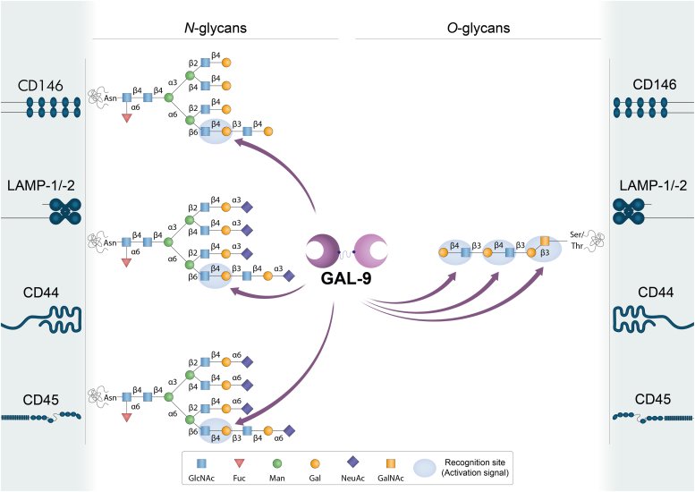
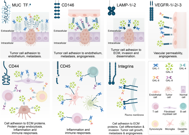
Galectins, a family of evolutionarily conserved glycan-binding proteins, play key roles in diverse biological processes including tissue repair, adipogenesis, immune cell homeostasis, angiogenesis, and pathogen recognition. Dysregulation of galectins and their ligands has been observed in a wide range of pathologic conditions including cancer, autoimmune inflammation, infection, fibrosis, and metabolic disorders. Through protein-glycan or protein-protein interactions, these endogenous lectins can shape the initiation, perpetuation, and resolution of these processes, suggesting their potential roles in disease monitoring and treatment. However, despite considerable progress, a full understanding of the biology and therapeutic potential of galectins has not been reached due to their diversity, multiplicity of cell targets, and receptor promiscuity. In this article, we discuss the multiple galectin-binding partners present in different cell types, focusing on their contributions to selected physiologic and pathologic settings. Understanding the molecular bases of galectin-ligand interactions, particularly their glycan-dependency, the biochemical nature of selected receptors, and underlying signaling events, might contribute to designing rational therapeutic strategies to control a broad range of pathologic conditions.

Keywords: angiogenesis; galectins; glycoproteins; glycosylation; immunity; receptors; tumorigenesis.


Conflict of interest statement

Conflict of interest M. F. T., M. T. E., A. G. B., L. S. and M. V. E. declare that they have no conflict of interest regarding the content of this article. G. A. R. reports a relationship with Galtec SAS that includes equities.

Figures

Figure 1
Figure 1
Conserved structuresof selected members of the galectin family and structural classification. Based on their structural features, galectins are classified into three groups: “proto-type” galectins (e.g. GAL-1) contain one carbohydrate recognition domain (CRD) and can dimerize; “tandem repeat-type” galectins (e.g. GAL-4, GAL-8 and GAL-9) contain two distinct CRD in tandem, connected by a linker peptide; and “chimera-type” GAL-3 which consists of unusual proline- and glycine-rich short stretches fused onto the CRD. GAL-1 structure is shown in blue (PDB: 4Y1U); GAL-3 structure in green (PDB: 4R9A); GAL-4 N-CRD in turquoise (PDB: 5DUV) and GAL-4 C-CRD in steel blue (PDB: 4YM3); protease-resistant mutant GAL-8 form possessing both N-CRD (shown in orange), and C-CRD (shown in red), with a linker of two amino acids (His-Met) (shown in green) (PDB: 3VKM); protease-resistant mutant GAL-9 form possessing both N-CRD (shown in lilac) and C-CRD (shown in violet) with a linker of 19 amino acids (shown in green) and a metal ion found at the CRDs interface (PDB: 3WV6). All structures are represented in complex with lactose in yellow and GAL-8 N-CRD with SiaLac in cyan.
Figure 2
Figure 2
Schematic representation of potential GAL-1-glycan interactions on selected cell surface receptors. GAL-1 preferentially recognizes terminal LacNAc residues on both N- and O-glycans. It can bind to terminal α2-3-linked sialic acid in the LacNAc sequence, but α2-6-linked sialic acid prevents the binding of this lectin. LacNAc: Galβ1-4GlcNAc. Glycans are represented according to the symbol nomenclature proposed (244).
Figure 3
Figure 3
Schematic representation of potential GAL-3-glycan interactions on selected cell surface receptors. GAL-3 preferentially binds to internal LacNAc residues on both N- and O-glycans. Terminal α2-3-linked or α2-6-linked sialic acid in the LacNAc sequence is permissive for binding of GAL-3, which can interact with internal LacNAc repeats. LacNAc: Galβ1-4GlcNAc. Glycans are represented according to the symbol nomenclature proposed (244).
Figure 4
Figure 4
Schematic representation of potential GAL-8-glycan interactions on selected cell surface receptors. GAL-8 binds to N- and O-glycans, and glycolipids. It preferentially recognizes internal -but also terminal- LacNAc residues. Sialyl-Lewis X tetrasaccharide (Neu5Acα2-3Galβ1-4[Fucα1-3]GlcNAcβ) may interact with GAL-8 in N- and O-glycans as well as in glycolipids. The N-terminal CRD of GAL-8 (GAL-8N) has a high binding affinity for α2-3-sialylated- or 3-sulfated β-galactosides and sialyl-Lewis X-containing N- or O-glycans and glycolipids. LacNAc: Galβ1-4GlcNAc. 3′Sialyl-LacNAc: Neu5Acα2-3Galβ1-4GlcNAc. 3′Sulfo-LacNAc: Sulfo-3Galβ1-4GlcNAc. Glycans are represented according to the symbol nomenclature proposed (244).
Figure 5
Figure 5
Schematic representation of GAL-9-glycan interactions on selected cell surface receptors. GAL-9 binds to N- and O-glycans. It preferentially recognizes internal LacNAc residues. The N-terminal CRD of GAL-9 (GAL-9N) has a high affinity for gangliosides (e.g. GA1, GM1, GD1a) and other glycolipids (ended in e.g. Förssman pentasaccharide, A-hexasaccharide). LacNAc: Galβ1-4GlcNAc. Glycans are represented according to the symbol nomenclature proposed (244).
Figure 6
Figure 6
Various glycosylated receptors are recognized by more than one galectin on different cell types. TF/MUC is a receptor for GAL-1, GAL-3, GAL-4, and GAL-8; CD146 is a glycosylated receptor for GAL-1, GAL-3, and GAL-9; LAMP-1/-2 is engaged by GAL-1 and GAL-3. N-glycan-dependent intracellular interaction between GAL-9 in the lysosomal lumen and LAMP-2 in gut epithelial cells (230) is not shown. VEGFR-1/-2/-3 and integrins are receptors for GAL-1, GAL-3 and GAL-8; CD44 and CD45 are receptors for GAL-1, GAL-3, GAL-8 and GAL-9. As a result of their glycan dependence or independence, the multivalent nature of these interactions, and the underlying signaling pathways, galectins play key roles in a wide range of biological programs.

References

    1. Yang R.Y., Rabinovich G.A., Liu F.T. Galectins: structure, function and therapeutic potential. Expert Rev. Mol. Med. 2008;10:1–24. - PubMed
    1. Thiemann S., Baum L.G. Galectins and immune responses-just how do they do those things they do? Annu. Rev. Immunol. 2016;34:243–264. - PubMed
    1. Kamili N.A., Arthur C.M., Gerner-Smidt C., Tafesse E., Blenda A., Dias-Baruffi M., et al. Key regulators of galectin-glycan interactions. Proteomics. 2016;16:3111–3125. - PMC - PubMed
    1. Hirabayashi J., Hashidate T., Arata Y., Nishi N., Nakamura T., Hirashima M., et al. Oligosaccharide specificity of galectins: a search by frontal affinity chromatography. Biochim. Biophys. Acta. 2002;1572:232–254. - PubMed
    1. Stowell S.R., Arthur C.M., Mehta P., Slanina K.A., Blixt O., Leffler H., et al. Galectin-1, -2, and -3 exhibit differential recognition of sialylated glycans and blood group antigens. J. Biol. Chem. 2008;283:10109–10123. - PMC - PubMed

Publication types