Review
. 2023 Oct 29;80(11):341.
doi: 10.1007/s00018-023-04998-z.

The role and mechanisms of microvascular damage in the ischemic myocardium

Affiliations
Review

The role and mechanisms of microvascular damage in the ischemic myocardium

Bang-Hao Zhao et al. Cell Mol Life Sci. .

Abstract

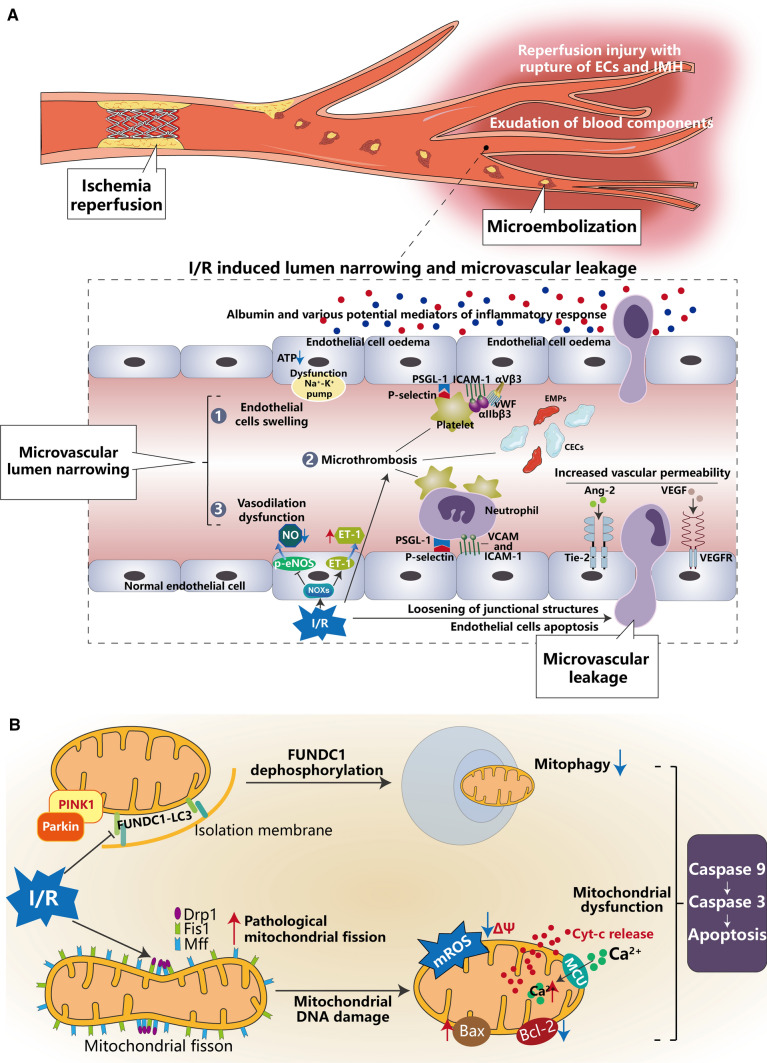
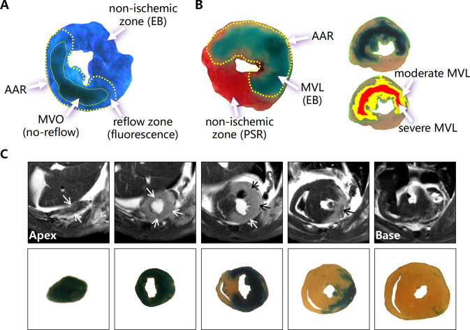
Following myocardial ischemic injury, the most effective clinical intervention is timely restoration of blood perfusion to ischemic but viable myocardium to reduce irreversible myocardial necrosis, limit infarct size, and prevent cardiac insufficiency. However, reperfusion itself may exacerbate cell death and myocardial injury, a process commonly referred to as ischemia/reperfusion (I/R) injury, which primarily involves cardiomyocytes and cardiac microvascular endothelial cells (CMECs) and is characterized by myocardial stunning, microvascular damage (MVD), reperfusion arrhythmia, and lethal reperfusion injury. MVD caused by I/R has been a neglected problem compared to myocardial injury. Clinically, the incidence of microvascular angina and/or no-reflow due to ineffective coronary perfusion accounts for 5-50% in patients after acute revascularization. MVD limiting drug diffusion into injured myocardium, is strongly associated with the development of heart failure. CMECs account for > 60% of the cardiac cellular components, and their role in myocardial I/R injury cannot be ignored. There are many studies on microvascular obstruction, but few studies on microvascular leakage, which may be mainly due to the lack of corresponding detection methods. In this review, we summarize the clinical manifestations, related mechanisms of MVD during myocardial I/R, laboratory and clinical examination means, as well as the research progress on potential therapies for MVD in recent years. Better understanding the characteristics and risk factors of MVD in patients after hemodynamic reconstruction is of great significance for managing MVD, preventing heart failure and improving patient prognosis.

Keywords: Microvascular damage; Microvascular endothelial cells; Microvascular leakage; Microvascular obstruction.


Conflict of interest statement

There are no conflicts of interest to declare.

Figures

Fig. 1
Fig. 1
Mechanisms of myocardial ischemia. (modified from [9] with a permission license 5,623,560,707,414) CFR coronary flow reserve, ACS acute coronary syndromes, AMI acute myocardial infarction, HT hypertension, SNS sympathetic nervous system, CMP cardiomyopathy
Fig. 2
Fig. 2
Pathological mechanisms of microvascular damage following myocardium ischemia/reperfusion
Fig. 3
Fig. 3
Simplified and incomplete scheme showing the molecular composition of endothelial junction complexes. Occludin, Claudin1/5, and ZO-1 constitute tight junction that are essential for endothelial barrier permeability. The central structural and functional component of adherens junction is VE–cadherin, which is uniquely expressed in ECs. Gap junction consist of connexin and allow the passage of molecules. Decrease in junction proteins and loosening of interendothelial cell junction structures due to ischemia/reperfusion are the main causes of leakage of blood components from microvasculature. ZO-1, zonula occludens protein-1
Fig. 4
Fig. 4
A Complex process of multifaceted and multifactorial interactions of microvascular damage in molecules, cellular and tissue levels after myocardial ischemia/reperfusion (I/R). After I/R injury, endothelial cells swelling, vasodilation dysfunction, and microthrombosis cause luminal narrowing, known as microvascular obstruction (MVO), whereas loosening of the interendothelial cell junction structures, massive death of endothelial cells and increased vascular permeability cause microvascular leakage (MVL). B Mitochondrial dysfunction leads to endothelial cells apoptosis. Due to I/R injury, mitochondrial fission/fusion was imbalanced and pathological mitochondrial fission damages mtDNA, promoting cytochrome c (cyt-c) release from mitochondria to the cytoplasm and activating the caspase family to promote endothelial cells apoptosis. Moreover, impaired mitophagy is unable to remove damaged mitochondrial debris, leading to a series of pathophysiological changes that eventually lead to endothelial cells injury and apoptosis. p-eNOS phosphorylated endothelial nitric oxide synthase, ET-1 endothelin-1, vWF von Willebrand Factor, PSGL-1 p-selectin glycoprotein ligand, ICAM intercellular adhesion molecules, CECs circulating endothelial cells, EMPs endothelial-derived microparticles, Ang-2 angiopoietin-2, Tie-2 TEK receptor tyrosine kinase a receptor of Ang-2, VEGF vascular endothelial growth factor, VEGFR vascular endothelial growth factor receptor, PINK1 PTEN-induced putative kinase, Parkin E3 ubiquitin ligase, FUNDC1 FUN14 domain-containing protein 1, LC3 light chain 3, Drp1 dynamin-related protein 1, Mff mitochondrial fission factor, Fis1 mitochondrial fission protein 1, mROS mitochondrial ROS, MCU the mitochondrial calcium uniporter complex
Fig. 5
Fig. 5
Experimental assays of microvascular damage after myocardial I/R injury. A Extent of MVO by the absence of thioflavin-S fluorescence (indicated by the thin yellow dotted line) within the AAR (indicated by the thick yellow dotted line) and NIZ by EB. B Left image detects MVL by EB within the AAR (indicated by the yellow dotted line) and NIZ by picrosirus red (PSR). The right image determines moderate MVL (yellow filled area) and severe MVL (red filled area). C Representative serial CMR images from the apex to the base of a mouse heart showing LGE (arrows) and serial cardiac transverse sections showing regions with MVL indicated by EB staining after I/R (1 h/24 h) (modified from [50])

References

    1. Li Y, Schoufour J, Wang DD, et al. Healthy lifestyle and life expectancy free of cancer, cardiovascular disease, and type 2 diabetes: prospective cohort study. BMJ. 2020;368:l6669. doi: 10.1136/bmj.l6669. - DOI - PMC - PubMed
    1. Bhatnagar A. Environmental determinants of cardiovascular disease. Circ Res. 2017;121(2):162–180. doi: 10.1161/CIRCRESAHA.117.306458. - DOI - PMC - PubMed
    1. Wang W, Hu M, Liu H, et al. Global Burden of Disease Study 2019 suggests that metabolic risk factors are the leading drivers of the burden of ischemic heart disease. Cell Metab. 2021;33(10):1943–1956e1942. doi: 10.1016/j.cmet.2021.08.005. - DOI - PubMed
    1. Balakumar P, Maung UK, Jagadeesh G. Prevalence and prevention of cardiovascular disease and diabetes mellitus. Pharmacol Res. 2016;113(Pt A):600–609. doi: 10.1016/j.phrs.2016.09.040. - DOI - PubMed
    1. Organization WH (2021) Cardiovascular diseases (CVDs). Retrieved from https://www.who.int/en/news-room/fact-sheets/detail/cardiovascular-disea...). Accessed 14 Feb 2023

MeSH terms