

RUNX1 rearrangement in mature B-cell acute lymphoblastic leukemia with non-L3 morphology
- PMID: 37899239
- PMCID: PMC10861373
- DOI: 10.3960/jslrt.23028
RUNX1 rearrangement in mature B-cell acute lymphoblastic leukemia with non-L3 morphology
Abstract
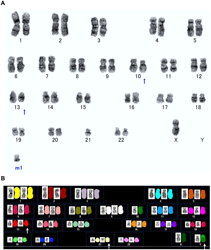
Mature B-cell acute lymphoblastic leukemia (ALL) is defined by the expression of light chain-restricted surface immunoglobulin (sIg) and usually has features of the leukemic phase of Burkitt lymphoma including FAB-L3 morphology and MYC rearrangement. Recently, another distinct entity in childhood mature B-cell ALL has been characterized as non-L3 morphology and KMT2A rearrangement. Here we report an unusual case of mature B-cell ALL that presented with RUNX1 rearrangement. A 65-year-old male was admitted to our department for thorough examination of leukocytosis and thrombocytopenia. The patient's bone marrow was hypercellular and infiltrated with 97.8% myeloperoxidase-negative, medium-to-large-sized blasts without cytoplasmic vacuoles. Immunophenotypes were characterized by the presence of light chain-restricted sIg and the lack of immature markers, indicating a diagnosis of mature B-cell ALL with L2 morphology: sIg-κ+, CD19+, CD20+, CD22+, CD79a+, TdT-, and CD34-. G-banding combined with spectral karyotyping showed the following complex karyotype: 45,X,der(Y;10)(p10;q10),del(13)(q?),inv(21)(p13q22.1). Fluorescence in situ hybridization revealed separated signals of RUNX1 at 21q22.1, whereas rearrangements of MYC and KMT2A were not found. To our knowledge, inv(21)(p13q22.1) involving RUNX1 is a novel cytogenetic aberration and this is the first case of mature B-cell ALL that presented with RUNX1 rearrangement. Thus, RUNX1 may be implicated in the pathogenesis of mature B-cell ALL showing non-L3 morphology without MYC rearrangement.
Keywords: RUNX1 rearrangement; fluorescence in situ hybridization; mature B-cell acute lymphoblastic leukemia; non-L3 morphology.
Conflict of interest statement
CONFLICT OF INTEREST
The authors declare no conflicts of interest.
Figures


References
-
- Bene MC, Castoldi G, Knapp W, et al. Proposals for the immunological classification of acute leukemias. European Group for the Immunological Characterization of Leukemias (EGIL). Leukemia. 1995; 9: 1783-1786. - PubMed
-
- Chan NPH, Ma ESK, Wan TSK, Chan LC. The spectrum of acute lymphoblastic leukemia with mature B-cell phenotype. Leuk Res. 2003; 27: 231-234. - PubMed
-
- Fukano R. Mature B-cell acute lymphoblastic leukemia. In : Kato M (ed) : Pediatric Acute Lymphoblastic Leukemia. Singapore, Springer. 2020; pp. 73-80.
-
- Leoncini L, Campo E, Stein H, et al. Burkitt lymphoma. In : Swerdlow SH, Campo E, Harris NL, et al. (eds) : WHO Classification of Tumors of Haematopoietic and Lymphoid Tissues. 4th ed, Lyon, IARC Press. 2017; pp. 330-334.
Publication types
MeSH terms
Substances
LinkOut - more resources
Full Text Sources
Research Materials
Miscellaneous