Comparative Study
. 2023 Oct 30;23(1):106.
doi: 10.1186/s40644-023-00626-y.

Head-to-head comparison of 18F-FAPI and 18F-FDG PET/CT in staging and therapeutic management of hepatocellular carcinoma

Affiliations
Comparative Study

Head-to-head comparison of 18F-FAPI and 18F-FDG PET/CT in staging and therapeutic management of hepatocellular carcinoma

Jing Zhang et al. Cancer Imaging. .

Abstract

Background: Fluorine 18 (18F) fluorodeoxyglucose (FDG) positron emission tomography/computed tomography (PET/CT) has limitations in staging hepatocellular carcinoma (HCC). The recently introduced 18F-labeled fibroblast-activation protein inhibitor (FAPI) has shown promising prospects in detection of HCC lesions. This study aimed to investigate the initial staging and restaging performance of 18F-FAPI PET/CT compared to 18F-FDG PET/CT in HCC.

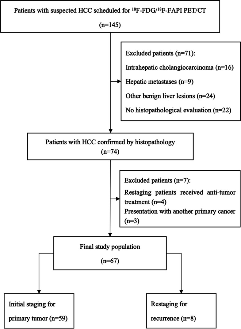
Methods: This prospective study enrolled histologically confirmed HCC patients from March 2021 to September 2022. All patients were examined with 18F-FDG PET/CT and 18F-FAPI PET/CT within 1 week. The maximum standard uptake value (SUVmax), tumor-to-background ratio (TBR), and diagnostic accuracy were compared between the two modalities.

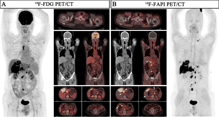
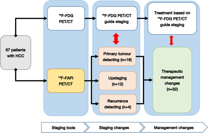
Results: A total of 67 patients (57 men; median age, 57 [range, 32-83] years old) were included. 18F-FAPI PET showed higher SUVmax and TBR values than 18F-FDG PET in the intrahepatic lesions (SUVmax: 6.7 vs. 4.3, P < 0.0001; TBR: 3.9 vs. 1.7, P < 0.0001). In diagnostic performance, 18F-FAPI PET/CT had higher detection rate than 18F-FDG PET/CT in intrahepatic lesions [92.2% (238/258) vs 41.1% (106/258), P < 0.0001] and lymph node metastases [97.9% (126/129) vs 89.1% (115/129), P = 0.01], comparable in distant metastases [63.6% (42/66) vs 69.7% (46/66), P > 0.05]. 18F-FAPI PET/CT detected primary tumors in 16 patients with negative 18F-FDG, upgraded T-stages in 12 patients and identified 4 true positive findings for local recurrence than 18F-FDG PET, leading to planning therapy changes in 47.8% (32/67) of patients.

Conclusions: 18F-FAPI PET/CT identified more primary lesions, lymph node metastases than 18F-FDG PET/CT in HCC, which is helpful to improve the clinical management of HCC patients.

Trial registration: Clinical Trials, NCT05485792 . Registered 1 August 2022, Retrospectively registered.

Keywords: 18F-FAPI; PET; CT; Hepatocellular carcinoma, Fibroblast activation protein.


Conflict of interest statement

The authors declare no conflict of interest.

Figures

Fig. 1
Fig. 1
Study flowchart shows inclusion and exclusion criteria. HCC = hepatocellular carcinoma; 18F = fluorine 18; FAPI = fibroblast activation protein inhibitor; FDG = fluorodeoxyglucose; PET/CT = positron emission tomography/computed tomography
Fig. 2
Fig. 2
Nine representative patients with HCC underwent 18F-FDG & 18F-FAPI PET/CT imaging. 18F-FAPI PET/CT outperformed.18F-FDG PET/CT in detecting primary tumors (Patient No. 32, 33, 34, 49), intrahepatic subfoci (Patient No. 13, 38, 42, 47, 48), supraclavicular lymph node metastases (Patient No. 13, 48), retroperitoneum lymph node metastases (Patient No. 48), and comparable in detecting distant metastases (Patient No. 48)
Fig. 3
Fig. 3
A 41-year-old male patient (Patient No. 51) with HCC (moderately differentiated) was confirmed by biopsy. 18F-FDG PET/CT displayed moderate uptake in the section II of the liver; However, the corresponding CT scan showed more nodules in other lobes of the liver. 18F-FAPI PET/CT detects greater radiotracer in primary lesions and other intrahepatic subfoci on both MIP (large arrow) and axial images (small arrow)
Fig. 4
Fig. 4
A 57-year-old male patient (Patient No. 50) with recurrent HCC (moderately differentiated) was confirmed by postoperative pathology. 18F-FDG PET/CT displayed no uptake in this lesion, although the corresponding CT scan showed lamellar low-density shadow in right lobe of the liver. 18F-FAPI PET/CT revealed intense uptake (SUVmax 9.0; TBR 6.0) in the recurrent lesion on both maximum intensity projection (MIP) (large arrow) and axial images (small arrow)
Fig. 5
Fig. 5
A 62-year-old male patient (Patient No. 13) with HCC (moderately differentiated) was confirmed by biopsy. Compared with 18F-FDG PET/CT, 18F-FAPI PET/CT revealed more intrahepatic subfoci (white arrow in axial images) and more lymph node metastases (red arrow in axial, coronal images). There was a lymph node in right upper mediastinum, showing low-uptake in 18F-FAPI but intense uptake in 18F-FDG, final pathological findings confirmed inflammatory
Fig. 6
Fig. 6
Overview of impact of 18F-FAPI PET/CT on staging and therapeutic management in HCC, therapeutic management was altered in 31 of 67 individuals

Similar articles

Cited by

References

    1. Villanueva A. Hepatocellular carcinoma. N Engl J Med. 2019;380:1450–1462. doi: 10.1056/NEJMra1713263. - DOI - PubMed
    1. Kesler M, Levine C, Hershkovitz D, et al. (68)Ga-PSMA is a novel PET-CT tracer for imaging of hepatocellular carcinoma: A prospective pilot study. J Nucl Med. 2019;60:185–191. doi: 10.2967/jnumed.118.214833. - DOI - PubMed
    1. Shi X, Xing H, Yang X, et al. Comparison of PET imaging of activated fibroblasts and (18)F-FDG for diagnosis of primary hepatic tumours: a prospective pilot study. Eur J Nucl Med Mol Imaging. 2021;48:1593–1603. doi: 10.1007/s00259-020-05070-9. - DOI - PubMed
    1. Hirmas N, Leyh C, Sraieb M, et al. (68)Ga-PSMA-11 PET/CT improves tumor detection and impacts management in patients with hepatocellular carcinoma. J Nucl Med. 2021;62:1235–1241. doi: 10.2967/jnumed.120.257915. - DOI - PMC - PubMed
    1. Park JW, Kim JH, Kim SK, et al. A prospective evaluation of 18F-FDG and 11C-acetate PET/CT for detection of primary and metastatic hepatocellular carcinoma. J Nucl Med. 2008;49:1912–1921. doi: 10.2967/jnumed.108.055087. - DOI - PubMed

Publication types

Associated data