Review
. 2023 Oct 30;17(1):451.
doi: 10.1186/s13256-023-04192-6.

Dedifferentiated endometrial carcinoma metastasis to axillary lymph node: a case report

Affiliations
Review

Dedifferentiated endometrial carcinoma metastasis to axillary lymph node: a case report

Chase William Morrison et al. J Med Case Rep. .

Abstract

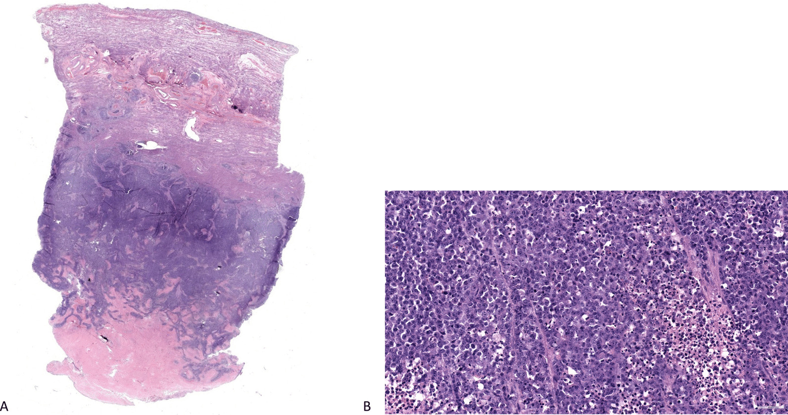
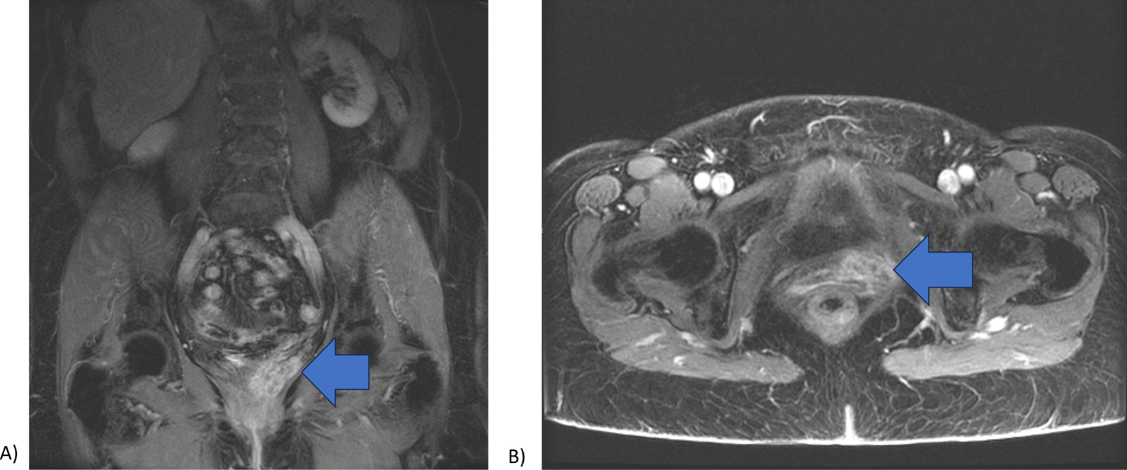
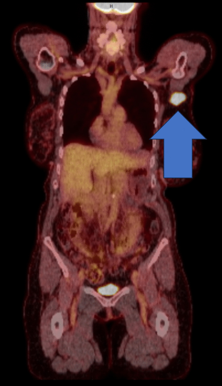
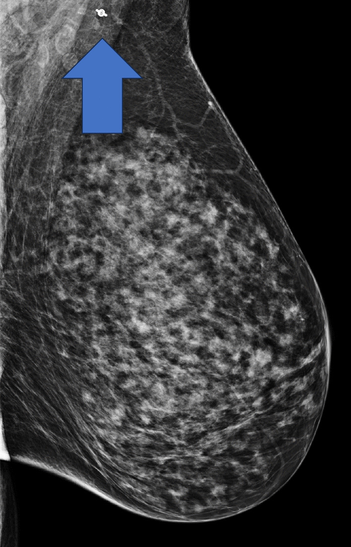
Background: We present an unusual case of a left axillary lymph node metastasis from a primary dedifferentiated endometrial carcinoma. This pattern of metastasis is likely the result of circulating tumor cells reaching the node through its arterial blood supply.

Case presentation: In this report, a 68-year-old white woman with a dedifferentiated endometrial carcinoma underwent a hysterectomy. She later developed an enlarged axillary lymph node due to metastatic dedifferentiated endometrial carcinoma, treated with chemotherapy and anti-programmed cell death protein 1 immunotherapy resulting in a complete clinical and radiological response.

Conclusion: A review of the literature reveals the rarity of blood-borne lymph node metastasis, especially with uterine carcinoma. Immunotherapy has shown promising results in the treatment of some subtypes of metastatic uterine carcinoma.

Keywords: Axillary mass; Case report; Endometrial carcinoma; Immunotherapy; Metastatic carcinoma.


Conflict of interest statement

The authors declare that they have no competing interests.

Figures

Fig. 1
Fig. 1
A Uterus, full thickness section, 2 ×, hematoxylin and eosin (H&E) stain; tumor extensively infiltrating myometrium (outer half) with lymphovascular invasion. B Uterus, 40 ×, H&E stain; tumor composed of diffuse sheets of loosely cohesive neoplastic cells with areas of necrosis; nuclear pleomorphism, prominent nucleoli, and brisk mitoses are seen, consistent with dedifferentiated component of endometrial carcinoma
Fig. 2
Fig. 2
a Heterogeneously enhancing structure, suggesting recurrence, found at the anterior vaginal cuff on surveillance CT abdomen and pelvis status post-hysterectomy (arrow) b Transverse section of CT pelvis illustrating anterior vaginal cuff recurrence (arrow)
Fig. 3
Fig. 3
a Mammography of the left breast showing axillary LN enhancement (arrow) b Targeted ultrasound of the left breast showing hypoechoic regions in sagittal and transverse planes (arrows)
Fig. 4
Fig. 4
PET-CT showing intense hypermetabolic region in the left axillary LN, arrow illustrating metastatic endometrial carcinoma
Fig. 5
Fig. 5
A Axillary lymph node 4 ×; H&E stain: needle core biopsy with areas of viable tumor (left) and extensive necrosis (right) B Axillary lymph node 40 ×; H&E stain: tumor composed of loosely cohesive neoplastic cells with pleomorphic nuclei and prominent nucleoli brisk mitosis, similar to the dedifferentiated component of primary endometrial carcinoma
Fig. 6
Fig. 6
Mammogram status post-pembrolizumab with arrow showing resolution of mass where the metallic biopsy marker is present

Similar articles

Cited by

References

    1. Kilinc ECT, Korkmaz V, Yalcin HR. Factors affecting lymph node metastasis in uterine papillary serous carcinomas: a retrospective analysis. J Obstets Gynecol. 2022;42:3725–3730. doi: 10.1080/01443615.2022.2158311. - DOI - PubMed
    1. Virchow R. Cellular pathology. Philadelphia: JB Lippincott; 1863.
    1. Tran BN, Grigsby PW, Dehdashti F, Herzog TJ, Siegel BA. Occult supraclavicular lymph node metastasis identified by FDG-PET in patients with carcinoma of the uterine cervix. Gynecol Oncol. 2003;90(3):572–576. doi: 10.1016/s0090-8258(03)00402-5. - DOI - PubMed
    1. Mochizuki Y, Tsuchiya M, Oyama J, Wada A, et al. Left supraclavicular (Virchow’s) node metastasis detected before primary infra-diaphragmatic tumor: a case series. J Med Case Rep. 2022;16:33. doi: 10.1186/s13256-022-03261-6. - DOI - PMC - PubMed
    1. Zhu Q, Li L, Jiao X, Xiong J, Zhai S, Zhu G, Cheng P, Qu J. Rare metastasis of gastric cancer to an axillary lymph node: a case report. Front Oncol. 2022;12:995738. doi: 10.3389/fonc.2022.995738. - DOI - PMC - PubMed