. 2023 Nov 1;31(6):682-691.
doi: 10.4062/biomolther.2023.162.

Licochalcone D Inhibits Skin Epidermal Cells Transformation through the Regulation of AKT Signaling Pathways

Affiliations

Licochalcone D Inhibits Skin Epidermal Cells Transformation through the Regulation of AKT Signaling Pathways

Sun-Young Hwang et al. Biomol Ther (Seoul). .

Abstract

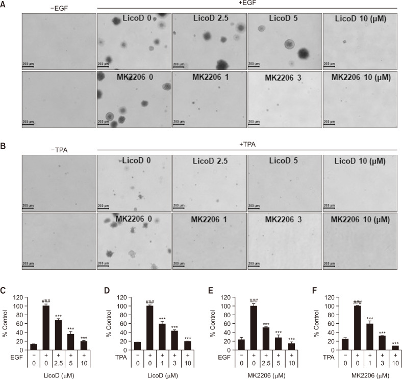
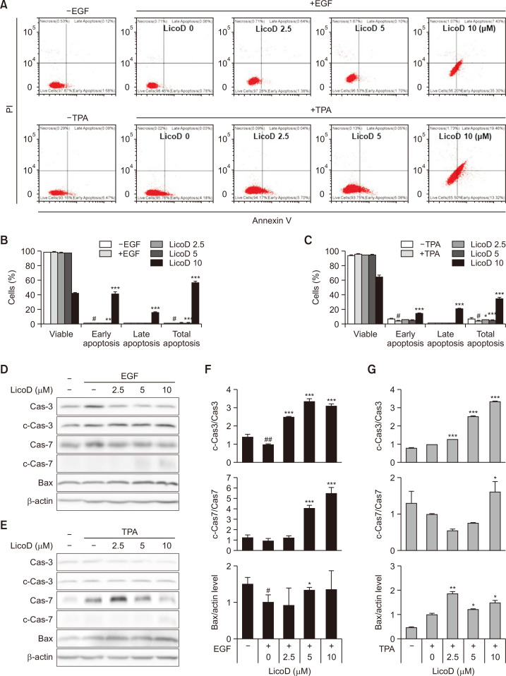
Cell transformation induced by epidermal growth factor (EGF) and 12-O-tetradecanoylphorbol-13-acetate (TPA) is a critical event in cancer initiation and progression, and understanding the underlying mechanisms is essential for the development of new therapeutic strategies. Licorice extract contains various bioactive compounds, which have been reported to have anticancer and anti-inflammatory effects. This study investigated the cancer preventive efficacy of licochalcone D (LicoD), a chalcone derivative in licorice extract, in EGF and TPA-induced transformed skin keratinocyte cells. LicoD effectively suppressed EGF-induced cell proliferation and anchorage-independent colony growth. EGF and TPA promoted the S phase of cell cycle, while LicoD treatment caused G1 phase arrest and down-regulated cyclin D1 and up-regulated p21 expression associated with the G1 phase. LicoD also induced apoptosis and increased apoptosis-related proteins such as cleaved-caspase-3, cleaved-caspase-7, and Bax (Bcl-2-associated X protein). We further investigated the effect of LicoD on the AKT signaling pathway involved in various cellular processes and found decreased p-AKT, p-GSK3β, and p-NFκB expression. Treatment with MK-2206, an AKT pharmacological inhibitor, suppressed EGF-induced cell proliferation and transformed colony growth. In conclusion, this study demonstrated the potential of LicoD as a preventive agent for skin carcinogenesis.

Keywords: AKT Signaling; Cell Transformation; EGF; Licochalcone D (LicoD); TPA.


Conflict of interest statement

CONFLICT OF INTEREST

The authors have no financial conflicts of interest to declare.

Figures

Fig. 1
Fig. 1
LicoD structure and its effects on cell viability on EGF- or TPA-induced cell proliferation. (A) The structure of LicoD, a natural com-pound isolated from licorice root, is shown. (B) Inhibition of EGF-induced JB6 cell proliferation by LicoD. EGF was treated at a concentration of 10 ng/mL, and various concentrations of LicoD were applied for 24 and 48 h. Statistical analysis was performed to evaluate the signifi-cance of the results, and any differences were considered statistically significant when p<0.05. (C) The morphological changes in EGF-induced cells by LicoD. The cells were treated with EGF (10 ng/mL) at various concentrations of LicoD (2.5, 5, 10 μM) in serum-free MEM for 48 h. (D) MK2206 inhibited EGF-induced JB6 cell proliferation. EGF (10ng/mL) and MK2206 (0, 1, 3, and 10 μM) were treated and incubated for 24 and 48 h. (E) The effect of MK2206 on morphological changes in EGF-induced transformed cells. JB6 cells were treated with EGF with 0, 1, 3, and 10 μM MK2206 and then incubated for 48 h. The morphological changes in the cells were observed using a microscope. Statistical analysis was performed to evaluate the significance of the results, and significantly different at: ###p<0.001 compared to the control; ***p<0.001 compared to group treated with EGF only.
Fig. 2
Fig. 2
LicoD and MK2206 suppress EGF- or TPA-induced JB6 cell transformation in soft agar. (A) Anchorage-independent cell growth was assessed by conducting soft agar assays with various concentrations of LicoD or MK2206 in combination with EGF. (B) Additionally, TPA and various concentrations of LicoD or MK2206 were co-treated to evaluate anchorage-independent cell growth. (C, D) The bar represents the relative ratio of colonies in soft agar with EGF or TPA and LicoD represented as the means ± SD of three replicates. (E, F) The bar graph indicates the relative ratio of colonies in soft agar with EGF or TPA and MK2206 represented as the means ± SD of three replicates. Significantly different at: ###p<0.001 compared to the control; ***p<0.001 compared to group treated with EGF or TPA only.
Fig. 3
Fig. 3
LicoD induces cell cycle arrest in EGF- or TPA-induced JB6 cells. (A) Flow cytometry analysis of EGF or TPA-induced JB6 cells treated with different concentrations of LicoD for 18 h. The representative images are shown. (B, C) The cell cycle distribution measured with a flowcytometry was represented as bar graphs. The effect of LicoD on the expression of proteins associated with cell cycle in EGF- (D, E) or TPA- (F, G) induced JB6 cells was investigated. JB6 cells were treated with EGF or TPA with 0, 2.5, 5, and 10 μM LicoD. The cell lysates were performed to western blotting using CyclinD1 and p21 antibodies. β-actin was used as a loading control. Densitometry quantification of proteins compared to β-actin is represented as the means ± SD. Significantly different at: ###p<0.001 compared to the control; **p<0.01 and ***p<0.001 compared to group treated with EGF or TPA only.
Fig. 4
Fig. 4
Lico D affects apoptosis in EGF- or TPA-induced JB6 cells. (A) The representative images of flow cytometry analysis in EGF- or TPA- induced JB6 cells treated with LicoD (2.5, 5, 10 μM) for 48 h. (B, C) The proportion of cells by stained with Annexin V/propidium iodide was represented as bar graphs. (D, E) Representative Western blots of apoptosis-related proteins expression of Caspase-3, Caspase-7 and Bax in EGF- or TPA-induced JB6 cells. JB6 cells were treated with EGF or TPA with 0, 2.5, 5, and 10 μM LicoD. The cell lysates were performed to western blotting using Caspase-3, Caspase-7 and Bax antibodies. β-actin was used as a loading control. (F, G) Quantitation of band intensity from blots by densitometry. The relative expression of cleaved-Caspase-3, cleaved-Caspase-7 and Bax was shown after normalization against Caspase-3, Caspase-7 and actin, respectively. Significantly different at: #p<0.05 and ##p<0.01 compared to the control; *p<0.05, **p<0.01, and ***p<0.001 compared to group treated with EGF or TPA only.
Fig. 5
Fig. 5
LicoD alters the expression of proteins associated with AKT signaling in EGF- or TPA-induced JB6 cells. (A) The effect of LicoD on the expression of AKT signaling-related proteins in EGF-induced JB6 cells was investigated. JB6 cells were treated with EGF with 0, 2.5, 5, and 10 μM LicoD and then incubated for 30 min. The cell lysates were performed to western blotting using p-AKT, panAKT, p-GSK3β, GSK3β, p-NFκB, NFκB, p-mTOR and mTOR antibodies. β-actin was used as a loading control. (B) The relative expression of p-AKT, p-mTOR, p-GSK3β and p-NFκB in EGF-induced JB6 cells was shown after normalization against panAKT, mTOR, GSK3β, and NFκB, respectively. (C) The effect of LicoD on the expression of AKT signaling-related proteins in TPA-induced JB6 cells was examined. JB6 cells were treated with TPA with 0, 2.5, 5, and 10 μM LicoD and then incubated for 60 min. (D) The relative expression of p-AKT, p-mTOR, p-GSK3β and p-NFκB in TPA-induced JB6 cells was shown after normalization against panAKT, mTOR, GSK3β, and NFκB, respectively. Significantly different at: #p<0.05, ##p<0.01, and ###p<0.001 compared to the control; *p<0.05, **p<0.01, and ***p<0.001 compared to group treated with EGF or TPA only.

References

    1. Aggarwal V., Tuli H. S., Tania M., Srivastava S., Ritzer E. E., Pandey A., Aggarwal D., Barwal T. S., Jain A., Kaur G., Sak K., Varol M., Bishayee A. Molecular mechanisms of action of epigallocatechin gallate in cancer: recent trends and advancement. Semin. Cancer Biol. 2022;80:256–275. doi: 10.1016/j.semcancer.2020.05.011. - DOI - PubMed
    1. Chatterjee N., Alfaro-Moreno E. In vitro cell transformation assays: a valuable approach for carcinogenic potentiality assessment of nanomaterials. Int. J. Mol. Sci. 2023;24:8219. doi: 10.3390/ijms24098219.01874d595bd24dd780b77a336720b736 - DOI - PMC - PubMed
    1. Deng N., Qiao M., Li Y., Liang F., Li J., Liu Y. Anticancer effects of licochalcones: a review of the mechanisms. Front. Pharmacol. 2023;14:1074506. doi: 10.3389/fphar.2023.1074506.59ff39ea66704d3fbae198bf2fd7d1b8 - DOI - PMC - PubMed
    1. Dias M. C., Pinto D., Silva A. M. S. Plant flavonoids: chemical characteristics and biological activity. Molecules. 2021;26:5377. doi: 10.3390/molecules26175377.f9bb94ac5c824d499f9641eaebf5dedd - DOI - PMC - PubMed
    1. Fu X., Zhao R., Yoon G., Shim J. H., Choi B. Y., Yin F., Xu B., Laster K. V., Liu K., Dong Z., Lee M. H. 3-Deoxysappanchalcone inhibits skin cancer proliferation by regulating T-lymphokine-activated killer cell-originated protein kinase in vitro and in vivo. Front. Cell Dev. Biol. 2021;9:638174. doi: 10.3389/fcell.2021.638174.5904eced933f4537ae619a32a502f211 - DOI - PMC - PubMed