

Editorial
doi: 10.1159/000530730.
Epub 2023 Apr 18.
The 5th Edition of the World Health Organization Classification of Tumours of the Eye and Orbit
Affiliations
- PMID: 37900189
- PMCID: PMC10601864
- DOI: 10.1159/000530730
Item in Clipboard
Editorial
The 5th Edition of the World Health Organization Classification of Tumours of the Eye and Orbit
Ocul Oncol Pathol.
2023 Sep.
No abstract available
Conflict of interest statement
The authors have no conflicts of interest to declare.
Figures

Combined nevocellular and WNT-activated deep penetrating/plexiform melanocytoma (nevus) (DPN) of the conjunctiva. a Circumscribed variably pigmented nodule in plica semilunaris, associated with a feeder vessel. b DPN component of the lesion is composed of lightly pigmented spindle melanocytes with abundant cytoplasm (asterisk), associated with scattered darkly pigmented melanophages (arrowhead). Mostly amelanotic nevocellular nevus (arrow) is present in the periphery. The DPN component of the lesion is positive for HMB45 (red chromogen, c), nuclear cyclin D1 (brown chromogen, d), and cytoplasmic and nuclear beta-catenin (brown chromogen, e). The nevocellular nevoid component of the lesion is negative for these markers (arrows, c–e). No proliferative activity is highlighted with Ki-67 (f) [hematoxylin-eosin (b), HMB45 (c), cyclin D1 (d), beta-catenin (e), Ki-67 (f); all figures. ×200].

Adenosquamous carcinoma of the conjunctiva. a Leukoplakic mass in the inferior fornix. b The invasive adenocarcinoma component is composed of epithelial cells with focal intracytoplasmic mucin (arrow) arranged in glands, in a background of desmoplastic stroma. Intracytoplasmic and luminal mucin is highlighted with Alcian blue stain (inset). c Sheets of atypical goblet cells (arrow). d Squamous cell carcinoma component is composed of irregular islands and bands of squamous cells [hematoxylin-eosin (b–d); ×100 (b), ×200 (c, d)].

Squamous cell carcinoma with mucinous differentiation. a Irregular islands and bands of invasive nonkeratinizing squamous cell carcinoma with focal intracytoplasmic mucin (b), highlighted with Alcian blue stain (c) [hematoxylin-eosin. ×50 (b), ×400 (b, c)].

Endocrine mucin-producing sweat gland carcinoma of the eyelid. a Reddish dermal-based nodule in the lower eyelid along the eyelash line. b Expansile nests of cells in cribriform arrangement. c Cuboidal neoplastic cells have ovoid, mildly pleomorphic nuclei, inconspicuous nucleoli, and moderate amounts of eosinophilic cytoplasm. d Alcian blue stain highlights intracytoplasmic and extracellular mucin. e Strong diffuse nuclear expression of estrogen receptors and insulinoma-associated protein 1 (INSM1) (f) [hematoxylin-eosin (b, c), Alcian blue (d), ER (e), INSM1 (f), ×50 (b), ×200 (c–f)].
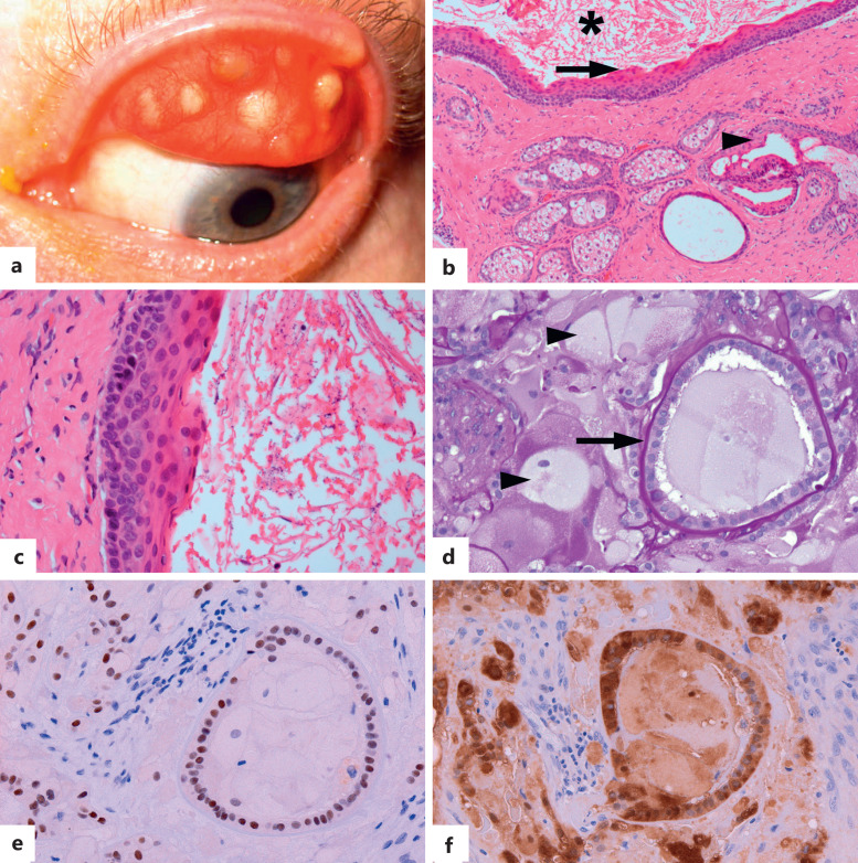
Tarsal cyst and phakomatous choristoma. a–c Tarsal cyst. a Tan-gray subepithelial tarsal-based nodules. b The cyst is lined by stratified squamous epithelium with eosinophilic corrugated delicately keratinized cuticle (arrow) and filled with keratin (asterisk). Meibomian gland lobules with associated ducts, lined by the epithelium morphologically similar to the cyst (arrowhead), are present in the adjacent tarsus. c Higher magnification of the cyst lining and delicate lamellated luminal keratin. d–f Phakomatous choristoma. d Cataractous-appearing tissue composed of bladder cells (arrowheads) and cuboidal lens epithelium associated with a prominent periodic acid-Schiff (PAS)-positive basement membrane, reminiscent of lens capsule (arrow). The lens epithelial cells express nuclear PAX8 (e) and cytoplasmic S100 (f) [hematoxylin-eosin (b, c), PAS (d), PAX8 (e), S100 (f), ×100 (b), ×1,000 (c), ×400 (c–f)].

MYCN-amplified retinoblastoma. a Gross photograph of the eye enucleated for failure to respond to local therapy demonstrates calcific post-treatment regression scar (arrow) and diffuse involvement of the detached retina by viable-appearing tumor (black arrowheads). Focal choroidal invasion is present (white arrowhead). Hemorrhage and necrosis are present in subretinal space (asterisk). b Histopathology demonstrates discohesive undifferentiated retinoblastoma cells, which focally invade choroid (arrow). c Discohesive retinoblastoma cells with prominent central nucleoli and scant cytoplasm (arrow), with brisk apoptotic bodies. d Retained retinoblastoma 1 (RB1) protein expression in the tumor [hematoxylin-eosin (b, c), RB1 (d), ×100 (b), ×400 (C), ×200 (d)]. Images courtesy of Ralph C. Eagle, Jr. MD.

Primary choroidal lymphoma (PCL), clinical and imaging findings. a Creamy choroidal infiltrates. b Optical coherence tomography demonstrates thickened choroid with an undulating hyperreflective anterior border (arrow) and obscuration of underlying choroidal vasculature from an infiltrative process.

Primary choroidal lymphoma (PCL), cytopathologic findings, fine needle aspiration biopsy. a Monomorphic small blue cells in a background of hemorrhage. Neoplastic cells express CD20 (b), bcl2 (c), and interferon regulatory factor 4 (IRF4/MUM1) (d). Neoplastic cells are negative for CD5, which highlights T cells (e) and CD10 (f). Overexpression of kappa light chain (g) over lambda (h). The Ki-67 labels rare nuclei (i) [hematoxylin-eosin (a), CD20 (b), bcl2 (c), MUM1 (d), CD5 (e), CD10 (f), kappa (g), lambda (h), ki-67 (i); all figures. ×400].

Primary vitreoretinal large B-cell lymphoma (PVR-LCBL), clinical and cytopathologic findings (subretinal fine needle aspiration biopsy and vitrectomy). a Punctate and nodular subretinal pigment epithelial (RPE) deposits (arrow), geographic placoid gray subretinal deposits focally associated with hemorrhage (black arrowhead), and areas of RPE atrophy. b Optical coherence tomography highlights subretinal (arrowhead) and sub-RPE (arrow) deposits. c Large cells with irregular nuclear contours, prominent nucleoli, and scant to moderate amounts of cytoplasm. d Foci of granulomatous inflammation (paraneoplastic granulomatous vitritis). e The large atypical cells and the apoptotic debris are positive for CD20 (e) and co-express bcl6 (f) and interferon regulatory factor 4 (IRF4/MUM1) (g). CD10 is negative (h). Ki-67 proliferative index is brisk (i). Targeted MYD88 mutation studies detected MYD88 p.L265P mutation [hematoxylin-eosin (c, d), CD20 (e), bcl6 (f), MUM1 (g), CD10 (h), Ki-67 (i), ×630 (c), ×400 (d–i)].
References
-
- International Agency for Research on Cancer [Internet]. Lyon: WHO Classification of Tumours Online. Available from: https://tumourclassification.iarc.who.int (accessed August 28, 2022).
-
- WHO classification of tumours editorial board. Eye tumours [Internet; beta version ahead of print]. WHO classification of tumours series. 5th ed.Lyon (France): International Agency for Research on Cancer; 2023. vol. 13 [cited]. Available from: https://tumourclassification.iarc.who.int/chapters/??.
-
- Grossniklaus HE, Eberhart CG, Kivelä TT. WHO classifiction of tumours of the eye. 4th ed.Lyon: International Agency for Research on Cancer; 2018.
-
- International Society for the Study of Vascular Anomalies [Internet] . 2018 ISSVA classification of vascular anomalies [cited 2018, may]. Available from: issva.org/classification.
Publication types
Grants and funding
LinkOut - more resources
Full Text Sources
