

In Vivo Kinematic Analysis of the Axial Shoulder Rotation in the Standing and Supine Positions Using 3D/2D Registration and Electromyography
- PMID: 37900413
- PMCID: PMC10613113
- DOI: 10.7759/cureus.46154
In Vivo Kinematic Analysis of the Axial Shoulder Rotation in the Standing and Supine Positions Using 3D/2D Registration and Electromyography
Abstract
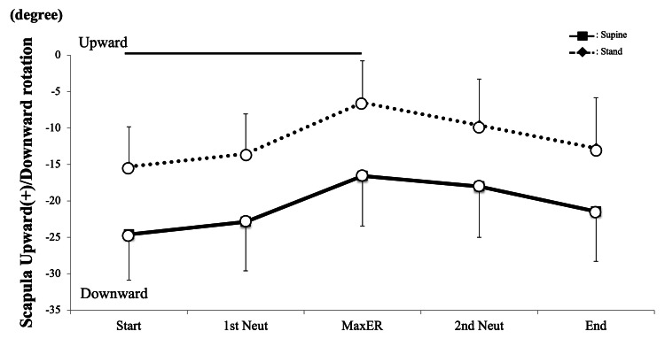
Background There has been no report comparing shoulder kinematics and muscle activities during axial shoulder rotation in different positions. The purpose of this study was to investigate differences in shoulder kinematics and muscle activities during axial shoulder rotation in healthy subjects between standing and supine positions using three-dimensional/two-dimensional (3D/2D) registration techniques and electromyography (EMG). Methods Eleven healthy males agreed to participate in this study. We recorded the fluoroscopy time during active shoulder axial rotation with a 90° elbow flexion in both standing and supine positions, simultaneously recording surface EMG of the infraspinatus, anterior deltoid, posterior deltoid, and biceps brachii. Three-dimensional bone models were created from CT images, and shoulder kinematics were analyzed using 3D/2D registration techniques. Muscle activities were evaluated as a ratio of mean electromyographic values to 5-sec maximum voluntary isometric contractions. Results Scapular kinematics during axial shoulder rotation in the supine position showed similar patterns with those in the standing position. The scapula was more posteriorly tilted and more downwardly rotated in the supine posture than in standing (P < 0.001 for both). Acromiohumeral distance (AHD) in the supine posture was significantly larger than in standing. Muscle activities showed no significant differences between postures except for biceps (P < 0.001). Discussion Shoulder kinematics and muscle activities during axial rotation were similar in pattern between standing and supine postures, but there were shifts in scapular pose and AHD. The findings of this study suggest that posture may be an important consideration for the prescription of optimal shoulder therapy following surgery or for the treatment of shoulder disorders.
Keywords: 3d/2d registration techniques; acromiohumeral distance; muscle activity; posture; scapula motion; shoulder rotation.
Copyright © 2023, Kenmoku et al.
Conflict of interest statement
The authors have declared that no competing interests exist.
Figures


Similar articles
-
Comparing in vivo three-dimensional shoulder elevation kinematics between standing and supine postures.JSES Int. 2021 Sep 4;5(6):1001-1007. doi: 10.1016/j.jseint.2021.07.005. eCollection 2021 Nov. JSES Int. 2021. PMID: 34766076 Free PMC article.
-
Electromyographic analysis of infraspinatus and scapular muscles during external shoulder rotation with different weight loads and positions.J Orthop Sci. 2019 Jan;24(1):75-80. doi: 10.1016/j.jos.2018.04.010. Epub 2018 Sep 6. J Orthop Sci. 2019. PMID: 30197094
-
Effect of different trunk postures on scapular muscle activities and kinematics during shoulder external rotation.J Shoulder Elbow Surg. 2019 Dec;28(12):2438-2446. doi: 10.1016/j.jse.2019.04.059. Epub 2019 Aug 10. J Shoulder Elbow Surg. 2019. PMID: 31409561
-
Shoulder muscle activity and function in common shoulder rehabilitation exercises.Sports Med. 2009;39(8):663-85. doi: 10.2165/00007256-200939080-00004. Sports Med. 2009. PMID: 19769415 Review.
-
Three-dimensional scapular orientation and muscle activity at selected positions of humeral elevation.J Orthop Sports Phys Ther. 1996 Aug;24(2):57-65. doi: 10.2519/jospt.1996.24.2.57. J Orthop Sports Phys Ther. 1996. PMID: 8832468 Review.
References
-
- Anterior shoulder stability: contributions of rotator cuff forces and the capsular ligaments in a cadaver model. Blasier RB, Guldberg RE, Rothman ED. J Shoulder Elbow Surg. 1992;1:140–150. - PubMed
-
- Mechanisms of glenohumeral joint stability. Lippitt S, Matsen F. Clin Orthop Relat Res. 1993;291:20–28. - PubMed
-
- Normal and abnormal motion of the shoulder. Poppen NK, Walker PS. https://pubmed.ncbi.nlm.nih.gov/1254624/ J Bone Joint Surg Am. 1976;58:195–201. - PubMed
-
- Glenohumeral contact forces. Anglin C, Wyss UP, Pichora DR. Proc Inst Mech Eng H. 2000;214:637–644. - PubMed
-
- The relevance of the moment arm of shoulder muscles with respect to axial rotation of the glenohumeral joint in four positions. Kuechle DK, Newman SR, Itoi E, Niebur GL, Morrey BF, An KN. Clin Biomech (Bristol, Avon) 2000;15:322–329. - PubMed
LinkOut - more resources
Full Text Sources