Clinical features and pregnancy outcomes in women with aortopulmonary window defect: case series
- PMID: 37900666
- PMCID: PMC10612472
- DOI: 10.1093/ehjcr/ytad501
Clinical features and pregnancy outcomes in women with aortopulmonary window defect: case series
Abstract
Background: Aortopulmonary window is a rare congenital heart defect that results in severe pulmonary arterial hypertension (PAH), Eisenmenger syndrome, and congestive heart failure in the first months of life. Pregnancy is absolutely contraindicated in the patients with this condition.
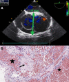
Case summary: This paper describes two clinical cases of pregnancy in patients (28 and 20 years old) with aortopulmonary window defect, severe PAH, and Eisenmenger syndrome that ended in preterm delivery by caesarean section. One patient died in the postpartum period due to progression of right ventricular heart failure. The younger patient survived childbirth and the postpartum period; later, she continued therapy at the PAH centre.
Discussion: We describe unusual cases of clinical features in pregnant women with aortopulmonary window defect. Due to the rare occurrence and low survival rate of patients with uncorrected aortopulmonary window defect, descriptions of clinical cases of this defect in adults are very rare. It is very important to note the necessity of observation of these patients in specialized centres by a multidisciplinary team of healthcare professionals, due to the high risk of cardiovascular, obstetric complications, and death.
Keywords: Aortopulmonary window; Case report; Eisenmenger syndrome; Pregnancy.
© The Author(s) 2023. Published by Oxford University Press on behalf of the European Society of Cardiology.
Conflict of interest statement
Conflict of interest: None declared.
Figures


References
-
- Barnes ME, Mitchell ME, Tweddell JS. Aortopulmonary window. Semin Thorac Cardiovasc Surg Pediatr Card Surg Annu 2011;14:67–74. - PubMed
-
- Umapathi KK, Nguyen H. Aortopulmonary window In: Statpearls [Internet]. Treasure Island, FL: StatPearls; 2021.
-
- Tkebuchava T, von Segesser LK, Vogt PR, Bauersfeld U, Jenni R, Künzli A, et al. . Congenital aortopulumonary window: diagnosis, surgical technique and long-term results. Eur J Cardiothorac Surg 1997;11:293–297. - PubMed
-
- Gowda D, Gajjar T, Rao JN, Chavali P, Sirohi A, Pandarinathan N, et al. . Surgical management of aortopulmonary window: 24 years of experience and lessons learned. Interact Cardiovasc Thorac Surg 2017;25:302–309. - PubMed
-
- Regitz-Zagrosek V, Roos-Hesselink JW, Bauersachs J, Blomström-Lundqvist C, Cifkova R, De Bonis M, et al. . 2018 ESC guidelines for the management cof cardiovascular diseases during pregnancy. Eur Heart J 2018;39:3165–3241. - PubMed
Publication types
LinkOut - more resources
Full Text Sources
