. 2023 Oct 24:17:3169-3192.
doi: 10.2147/DDDT.S420135. eCollection 2023.

Network Pharmacology Analysis and Machine-Learning Models Confirmed the Ability of YiShen HuoXue Decoction to Alleviate Renal Fibrosis by Inhibiting Pyroptosis

Affiliations

Network Pharmacology Analysis and Machine-Learning Models Confirmed the Ability of YiShen HuoXue Decoction to Alleviate Renal Fibrosis by Inhibiting Pyroptosis

MinChao Feng et al. Drug Des Devel Ther. .

Abstract

Purpose: YiShen HuoXue decoction (YSHXD) is a formulation that has been used clinically for the treatment of renal fibrosis (RF) for many years. We aimed to clarify therapeutic effects of YSHXD against RF and potential pharmacological mechanisms.

Materials and methods: We used network pharmacology analysis and machine-learning to screen the core components and core targets of YSHXD against RF, followed by molecular docking and molecular dynamics simulations to confirm the reliability of the results. Finally, we validated the network pharmacology analysis experimentally in HK-2 cells and a rat model of RF established by unilateral ureteral ligation (UUO).

Results: Quercetin, kaempferol, luteolin, beta-sitosterol, wogonin, stigmasterol, isorhamnetin, baicalein, and dihydrotanshinlactone progesterone were identified as the main active components of YSHXD in the treatment of unilateral ureteral ligation-induced RF, with IL-6, IL1β, TNF, AR, and PTGS2 as core target proteins. Molecular docking and molecular dynamics simulations further confirmed the relationship between compounds and target proteins. The potential molecular mechanism of YSHXD predicted by network pharmacology analysis was confirmed in HK-2 cells and UUO rats. YSHXD downregulated NLRP3, ASC, NF-κBp65, Caspase-1, GSDMD, PTGS2, IL-1β, IL-6, IL-18, TNF-α, α-SMA and upregulated HGF, effectively alleviating the RF process.

Conclusion: YSHXD exerts important anti-inflammatory and anti-cellular inflammatory necrosis effects by inhibiting the NLRP3/caspase-1/GSDMD-mediated pyroptosis pathway, indicating that YSHXD represents a new strategy and complementary approach to RF therapy.

Keywords: YiShen HuoXue decoction; machine-learning; molecular docking simulation; pyroptosis; renal fibrosis.


Conflict of interest statement

The authors declare that there are no conflicts of interest in this work.

Figures

None
Graphical abstract
Figure 1
Figure 1
Network pharmacology target analysis. (a) UpSet diagram of the three component databases; (b) Venn diagram of five disease target databases; (c) Venn diagram of the targets of the active components of YSHXD and the RF-related targets; (d) 56 overlapping YSHXD target PPI network diagram; (e) 22 overlapping YSHXD-RF target PPI network diagram.
Figure 2
Figure 2
Enrichment analysis of network pharmacological targets and multidimensional network graph analysis. (a) GO functional enrichment analysis of the 56 overlapping YSHXD targets; (b) KEGG enrichment analysis of the 56 overlapping YSHXD targets; (c) Bubble chart of the KEGG signaling pathways associated with YSHXD; (d) Bubble chart of the KEGG signaling pathways associated with the therapeutic effect of YSHXD on RF; (e) Herb-active component-target network diagram; (f) Sankey diagram of the herb-main active component-target-pathway.
Figure 3
Figure 3
Partial results of molecular docking and molecular dynamics simulation of YSHXD core active components and proteins. (a) AR-luteolin; (b) PTGS2-beta-sitosterol; (c) IL1B-luteolin; (d) TNF-luteolin; (e) IL6-kaempferol, (f) heat map of LibDock scores and (g) RMSD, RMSF and Rg of key active components and target proteins of YSHXD.
Figure 4
Figure 4
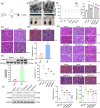
Effect of UUO by surgical ligation on renal tissues and effects of YSHXD on the general and histopathological characteristics of UUO model rats. (a) Experimental design scheme; (b) diagram of the kidney appearance (n = 6/group); (c) rat renal histopathology by H&E staining (n = 6/group) (× 200 and × 400 magnification); (d) Masson’s trichrome staining (n = 6/group, paired two-tailed t-test) (× 400 magnification); (e) representative Western blot images of α-SMA expression (n = 6/group, paired two-tailed t-test). (f) Body weight (n = 6/group, two‑way ANOVA with Sidak’s post-hoc test); (g) renal index (n = 6/group, one‑way ANOVA with Bonferroni post-hoc test); (h) H&E staining (n = 6/group) (× 200 and × 400 magnification); (i) Masson’s trichrome staining (× 400 magnification) and collagen fiber area (%) (n = 6/group, one‑way ANOVA with Bonferroni post-hoc test); (j) representative Western blot images of α-SMA and PTGS2 levels (n = 6/group, one‑way ANOVA with Bonferroni post-hoc test). Data were represented as the mean ± SD. ****P < 0.0001 vs Sham group; ##P < 0. 01 and ####P < 0.0001 vs Model group.
Figure 5
Figure 5
YSHXD protects renal tissues in UUO model rats (n = 6) by regulating pyroptosis. (a) Serum levels of inflammatory factors in UUO rats measured by ELISA (n = 6/group, one‑way ANOVA with Bonferroni post-hoc test); (b) representative immunohistochemical staining of NLRP3, caspase-1, GSDMD, and IL-1β in renal tissues (× 400 magnification); (c) quantification of the data shown in (b) (n = 6/group, one‑way ANOVA with Bonferroni post-hoc test); (d) protein expression levels of NLRP3, ASC, NF-κB, caspase-1, GSDMD, IL-18, IL-1β, and IL-6 determined by immunoblotting; (e) quantification of the data shown in (d) (n = 6/group, one‑way ANOVA with Bonferroni post-hoc test). Data were represented as the mean ± SD. ****P < 0.0001 vs Sham group; ###P < 0.001 and ####P < 0.0001 vs Model group.
Figure 6
Figure 6
YSHXD inhibited pyroptosis and exerted anti-RF effects in HIK-2 cells. (a) Viability of HK-2 cells measured by CCK-8 assay (n = 6/group, one‑way ANOVA with Bonferroni post-hoc test) and images of HK-2 cell morphology (× 200 magnification); (b) levels of IL-1β, IL-6, IL-8, IL-18, TNF-α and HGF in HK-2 cell culture supernatants measured by ELISA (n = 6/group, one‑way ANOVA with Bonferroni post-hoc test); (c) transcript levels of NLRP3, NF-κB, caspase-1, GSDMD, IL-1β, and IL-6 in TGF-β-induced HK-2 cells measured by RT-qPCR (n = 6/group, Welch ANOVA with Dunnett’s T3 post-hoc test); (d) protein levels of NLRP3, NF-κB, caspase-1, GSDMD, IL-1β, and IL-6 determined by Western blotting; (e) quantification of the data shown in (d) (n = 6/group, one‑way ANOVA with Bonferroni post-hoc test). Data were represented as the mean ± SD. ****P < 0.0001 vs Control group; ##P < 0.01 and ####P < 0.0001 vs Model group.
Figure 7
Figure 7
Molecular docking and molecular dynamics simulation of core active components of YSHXD and proteins (partial results). (a) NLRP3-beta-sitosterol; (b) NF-κB-kaempferol; (c) caspase-1-quercetin; (d) GSDMD-luteolin; (e) IL-18-luteolin; (f) heat map of LibDock scores, and (g) RMSD, RMSF and hydrogen bond heat map of key active components and target proteins.
Figure 8
Figure 8
Schematic representation of the therapeutic mechanism of YSHXD in renal fibrosis. In UUO or TGF-β-induced fibrosis models, pro-inflammatory cytokine levels were raised, pyroptosis was activated, and inflammatory factors were released, leading to renal fibrosis. YSHXD inhibits pyroptosis by inhibiting NLRP3/caspase-1/GSDMD activation to alleviate renal fibrosis.

References

    1. Wang Y-N, Ma S-X, Chen -Y-Y, et al. Chronic kidney disease: biomarker diagnosis to therapeutic targets. Clin Chim Acta. 2019;499:54–63. doi:10.1016/j.cca.2019.08.030 - DOI - PubMed
    1. Naber T, Purohit S. Chronic Kidney Disease: role of Diet for a Reduction in the Severity of the Disease. Nutrients. 2021;13(9):57. - PMC - PubMed
    1. Humphreys BD. Mechanisms of Renal Fibrosis. Annu Rev Physiol. 2018;80:309–326. doi:10.1146/annurev-physiol-022516-034227 - DOI - PubMed
    1. Hou FF, Lu YH. New insights into the pathogenesis and therapeutics of kidney fibrosis. Kidney Int Suppl. 2014;4(1):1. doi:10.1038/kisup.2014.1 - DOI - PMC - PubMed
    1. Higgins SP, Tang Y, Higgins CE, et al. TGF-beta1/p53 signaling in renal fibrogenesis. Cell Signal. 2018;43:1–10. doi:10.1016/j.cellsig.2017.11.005 - DOI - PMC - PubMed