. 2023 Sep 26;15(9):876-896.
doi: 10.4252/wjsc.v15.i9.876.

Interferon-γ priming enhances the therapeutic effects of menstrual blood-derived stromal cells in a mouse liver ischemia-reperfusion model

Affiliations

Interferon-γ priming enhances the therapeutic effects of menstrual blood-derived stromal cells in a mouse liver ischemia-reperfusion model

Qi Zhang et al. World J Stem Cells. .

Abstract

Background: Mesenchymal stem cells (MSCs) have been used in liver transplantation and have certain effects in alleviating liver ischemia-reperfusion injury (IRI) and regulating immune rejection. However, some studies have indicated that the effects of MSCs are not very significant. Therefore, approaches that enable MSCs to exert significant and stable therapeutic effects are worth further study.

Aim: To enhance the therapeutic potential of human menstrual blood-derived stromal cells (MenSCs) in the mouse liver ischemia-reperfusion (I/R) model via interferon-γ (IFN-γ) priming.

Methods: Apoptosis was analyzed by flow cytometry to evaluate the safety of IFN-γ priming, and indoleamine 2,3-dioxygenase (IDO) levels were measured by quantitative real-time reverse transcription polymerase chain reaction, western blotting, and ELISA to evaluate the efficacy of IFN-γ priming. In vivo, the liver I/R model was established in male C57/BL mice, hematoxylin and eosin and TUNEL staining was performed and serum liver enzyme levels were measured to assess the degree of liver injury, and regulatory T cell (Treg) numbers in spleens were determined by flow cytometry to assess immune tolerance potential. Metabolomics analysis was conducted to elucidate the potential mechanism underlying the regulatory effects of primed MenSCs. In vitro, we established a hypoxia/reoxygenation (H/R) model and analyzed apoptosis by flow cytometry to investigate the mechanism through which primed MenSCs inhibit apoptosis. Transmission electron microscopy, western blotting, and immunofluorescence were used to analyze autophagy levels.

Results: IFN-γ-primed MenSCs secreted higher levels of IDO, attenuated liver injury, and increased Treg numbers in the mouse spleens to greater degrees than untreated MenSCs. Metabolomics and autophagy analyses proved that primed MenSCs more strongly induced autophagy in the mouse livers. In the H/R model, autophagy inhibitors increased the level of H/R-induced apoptosis, indicating that autophagy exerted protective effects. In addition, primed MenSCs decreased the level of H/R-induced apoptosis via IDO and autophagy. Further rescue experiments proved that IDO enhanced the protective autophagy by inhibiting the mammalian target of rapamycin (mTOR) pathway and activating the AMPK pathway.

Conclusion: IFN-γ-primed MenSCs exerted better therapeutic effects in the liver I/R model by secreting higher IDO levels. MenSCs and IDO activated the AMPK-mTOR-autophagy axis to reduce IRI, and IDO increased Treg numbers in the spleen and enhanced the MenSC-mediated induction of immune tolerance. Our study suggests that IFN-γ-primed MenSCs may be a novel and superior MSC product for liver transplantation in the future.

Keywords: Autophagy; Cell therapy; Liver; Mesenchymal stem cells; Reperfusion injury; T-lymphocytes.


Conflict of interest statement

Conflict-of-interest statement: All the authors report no relevant conflicts of interest for this article.

Figures

Figure 1
Figure 1
Animal experiment grouping and timeline. Detailed descriptions are provided in the Materials and Methods. In brief, we established short-term and long-term liver ischemia-reperfusion mouse models to explore the therapeutic potential of menstrual blood-derived stromal cells (MenSCs) and primed MenSCs. MenSCs: Menstrual blood-derived stromal cells; IFN-γ: Interferon-γ; 1MT: 1-methyl-D-tryptophan; IR: Ischemia-reperfusion; IRM: Combination treated with ischemia-reperfusion and menstrual blood-derived stromal cells; IRM (IFN-γ): Combination treated with ischemia-reperfusion and interferon-γ-primed menstrual blood-derived stromal cells; PBS: Phosphate buffered saline; i.v.: Intravenous injection.
Figure 2
Figure 2
Characterization of menstrual blood-derived stromal cells and menstrual blood-derived stromal cells primed with interferon-γ. A: The surface marker expression of menstrual blood-derived stromal cells (MenSCs) was assessed by flow cytometry analysis; B: The osteogenic differentiation potential of MenSCs. Scale bar 200 μm; C: The chondrogenic differentiation potential of MenSCs. Scale bar 200 μm; D: The adipogenic differentiation potential of MenSCs. Scale bar 200 μm; E: MenSC apoptosis was detected by flow cytometry. The red box indicates the sum of late and early apoptotic cells, which represent the overall level of apoptosis; F: The overall level of apoptosis was analyzed; G: After MenSC priming with 100 ng/mL interferon-γ for 24 h, 48 h, 72 h, and 72 h + hypoxia/reoxygenation, the mRNA levels of indoleamine 2,3-dioxygenase (IDO) in MenSCs were determined by quantitative real-time reverse transcription polymerase chain reaction analysis; H: The protein level of IDO in MenSCs was determined by western blotting; I: The level of IDO secreted by MenSCs into the cell culture supernatant was measured by ELISA. The data are shown as the means ± SEMs, n = 3. aP < 0.05, bP < 0.01, cP < 0.001, dP < 0.0001. NS represents not statistically significant. All P values were obtained by one-way ANOVA. HLA-DR: Human leukocyte antigen-DR; IgG: Immunoglobulin G; NC: Negative control; HR: Hypoxia/reoxygenation; PI: Propidium iodide; IDO: Indoleamine 2,3-dioxygenase.
Figure 3
Figure 3
Interferon-γ-primed menstrual blood-derived stromal cells attenuated liver ischemia-reperfusion injury in the mouse short-term ischemia-reperfusion model. A: Representative images of histological liver sections stained with hematoxylin and eosin (hematoxylin and eosin staining, magnification 200 ×, images in the second row are partial magnifications of regions indicated by the red box); B: The degree of liver injury in each group was determined according to Suzuki’s injury score. The data are expressed as the means ± SEMs (n = 10/group); C: Representative images of liver sections from each group stained for caspase-3 to evaluate the apoptosis level (magnification 200 ×, images in the second row are partial magnifications of regions indicated by the red box); D: Representative liver sections from each group stained with TUNEL were obtained to evaluate the apoptosis level (magnification 200 ×, images in the second row are partial magnifications of regions indicated by the red box); E: The serum alanine aminotransferase levels were measured to indicate liver function levels, the data are expressed as the means ± SEMs (n = 3/group); F: The serum aspartate aminotransferase levels were measured to indicate liver function levels, and the data are expressed as the means ± SEMs (n = 3/group). aP < 0.05, bP < 0.01, cP < 0.001, dP < 0.0001. NS represents not statistically significant. All P values were obtained by one-way ANOVA. MenSCs: Menstrual blood-derived stromal cells; IFN-γ: Interferon-γ; IR: Ischemia-reperfusion; IRM: Combination treated with ischemia-reperfusion and menstrual blood-derived stromal cells; IRM (IFN-γ): Combination treated with ischemia-reperfusion and interferon-γ-primed menstrual blood-derived stromal cells; HE: Hematoxylin and eosin; TUNEL: Terminal deoxynucleotidyl transferase-mediated dUTP-biotin Nick End Labeling; ALT: Alanine aminotransferase; AST: Aspartate aminotransferase.
Figure 4
Figure 4
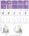
Interferon-γ-primed menstrual blood-derived stromal cells reduced hepatocellular damage and increased the Treg numbers in the mouse long-term ischemia-reperfusion model. A: Representative images of histological liver sections stained with hematoxylin and eosin were obtained (hematoxylin and eosin staining, magnification 200×, images in the second row are partial magnifications of regions indicated by the red box); B: The degree of liver injury in each group was determined according to Suzuki’s injury score. The data are expressed as the means ± SEMs (n = 3-5/group); C: The numbers of CD4, CD25, and FoxP3 positive cells (regulatory T cells) in the spleen were determined by flow cytometry to estimate the ability of primed menstrual blood-derived stromal cells to induce immune tolerance; D: The percentages of Tregs are expressed as the means ± SEMs (n = 3/group). aP < 0.05, bP < 0.01, cP < 0.001, dP < 0.0001. NS represents not statistically significant. All P values were obtained by one-way ANOVA. MenSCs: Menstrual blood-derived stromal cells; IFN-γ: Interferon-γ; 1MT: 1-methyl-D-tryptophan; IR: Ischemia-reperfusion; IRM: Combination treated with ischemia-reperfusion and menstrual blood-derived stromal cells; IRM (IFN-γ): Combination treated with ischemia-reperfusion and interferon-γ-primed menstrual blood-derived stromal cells; HE: Hematoxylin and eosin; Treg: Regulatory T cell.
Figure 5
Figure 5
Metabolomic data analysis. A: Heatmap of 45 differential metabolites between the ischemia-reperfusion (IR) group and IRM (interferon-γ) group; B: Volcano plot of 45 differential metabolites; C: The relevant pathways in which the differential metabolites are involved were analyzed by comparing the information in the Kyoto Encyclopedia of Genes and Genomes database; D: The main biological functions of differential metabolites were elucidated by pathway enrichment analysis; E: Relative quantitation of the differential metabolite phosphatidyl ethanolamine was performed to measure the effect of primed menstrual blood-derived stromal cells on the level of autophagy in liver tissues. MenSCs: Menstrual blood-derived stromal cells; IFN-γ: Interferon-γ; IR: Ischemia-reperfusion; IRM(IFN-γ) or IRPM: Combination treated with ischemia-reperfusion and interferon-γ-primed menstrual blood-derived stromal cells; PE: Phosphatidyl ethanolamine; OS: Organismal systems; M: Metabolism; HD: Human diseases; CP: Cellular processes; EIP: Environmental information processing; KEEG: Kyoto Encyclopedia of Genes and Genomes.
Figure 6
Figure 6
Interferon-γ-primed menstrual blood-derived stromal cells enhanced autophagy in mouse livers. A: Representative TEM images were used to determine the number of autophagic vacuoles in liver tissues from each experimental group. Scale bar 2 μm. The images in the second row are partial magnifications of the regions indicated by the red box; B: The number of autophagosomes was counted by TEM. The data are expressed as the means ± SEMs (n = 3/group); C: The expression of p-AMPK, AMPK, p-mammalian target of rapamycin (p-mTOR), mTOR, LC3, and ACTB in liver tissues was determined by western blotting; D: The relative normalized expression was quantified based on the intensities of the western blot bands (n = 3/group). The data are presented as the means ± SEMs. aP < 0.05, bP < 0.01, cP < 0.001, dP < 0.0001. NS represents not statistically significant. All P values were obtained by one-way ANOVA. MenSCs: Menstrual blood-derived stromal cells; IFN-γ: Interferon-γ; IR: Ischemia-reperfusion; IRM: Combination treated with ischemia-reperfusion and menstrual blood-derived stromal cells; IRM (IFN-γ): Combination treated with ischemia-reperfusion and interferon-γ-primed menstrual blood-derived stromal cells; TEM: Transmission electron microscopy; AMPK: The AMP-activated protein kinase signaling pathway; mTOR: Mammalian target of rapamycin; Tregs: Regulatory T cells.
Figure 7
Figure 7
interferon-γ-primed menstrual blood-derived stromal cells decreased hypoxia/reoxygenation-induced cell apoptosis via autophagy and indoleamine 2,3-dioxygenase. A: Apoptosis of L02 cells after different treatments was analyzed; B: The sum of the percentage of early apoptotic cells and late apoptotic cells was analyzed. The total percentage of apoptotic cells (sum of the percentage of early apoptotic cells and late apoptotic cells) was analyzed. The data are shown as the mean ± SEM, n = 3; bP < 0.01, dP < 0.0001. NS represents not statistically significant. All P values were obtained by one-way ANOVA. PI: Propidium iodide; NC: Negative control; 3MA: 3-methyladenine; 1MT: 1-methyl-D-tryptophan; MenSCs: Menstrual blood-derived stromal cells; IFN-γ: Interferon-γ; HR: Hypoxia/reoxygenation; HRM: Combination treated with hypoxia/reoxygenation and menstrual blood-derived stromal cells; HRM (IFN-γ): Combination treated with hypoxia/reoxygenation and I interferon-γ-primed menstrual blood-derived stromal cells.
Figure 8
Figure 8
Indoleamine 2,3-dioxygenase secreted by interferon-γ-primed menstrual blood-derived stromal cells enhanced autophagy by inhibiting the mammalian target of rapamycin pathway and activating the AMPK pathway. A: Immunofluorescence images of GFP-LC3. Scale bar 2 μm; B: The number of intense punctate GFP-LC3 aggregates in each cell was counted to determine the level of autophagy in each group. The data are expressed as the means ± SEMs (n = 10/group); C: The expression of p-AMPK, AMPK, p-mammalian target of rapamycin (p-mTOR), mTOR, LC3, and ACTB in L02 cells was determined by western blotting; D: The relative normalized protein expression was quantified based on the intensities of the western blot bands (n = 3/group). The data are presented as the means ± SEMs. aP < 0.05, bP < 0.01, cP < 0.001, dP < 0.0001. NS represents not statistically significant. All P values were obtained by one-way ANOVA. NC: Negative control; 3MA: 3-methyladenine; 1MT: 1-methyl-D-tryptophan; MenSCs: Menstrual blood-derived stromal cells; IFN-γ: Interferon-γ; HR: Hypoxia/reoxygenation; HRM: Combination treated with hypoxia/reoxygenation and menstrual blood-derived stromal cells; HRM (IFN-γ): Combination treated with hypoxia/reoxygenation and I interferon-γ-primed menstrual blood-derived stromal cells; AMPK: AMP-activated protein kinase; mTOR: the Mammalian target of rapamycin signaling pathway.
Figure 9
Figure 9
Schematic depiction of the mechanism. Ischemia-reperfusion injury (IRI) and immune rejection are two important factors that affect the prognosis of liver transplantation. After priming menstrual blood-derived stromal cells (MenSCs) with interferon-γ, the MenSCs secreted high levels of indoleamine 2,3-dioxygenase (IDO). On the one hand, MenSCs and IDO enhanced the AMPK-mammalian target of rapamycin-autophagy axis to reduce IRI; on the other hand, IDO promoted the differentiation of lymphocytes into Tregs to reduce immune rejection. IRI: Ischemia-reperfusion injury; MenSCs: Menstrual blood-derived stromal cells; IFN-γ: Interferon-γ; IDO: Indoleamine 2,3-dioxygenase; AMPK: AMP-activated protein kinase signaling pathway; mTOR: Mammalian target of rapamycin signaling pathway; Tregs: Regulatory T cells.

Similar articles

Cited by

References

    1. Nakano R, Tran LM, Geller DA, Macedo C, Metes DM, Thomson AW. Dendritic Cell-Mediated Regulation of Liver Ischemia-Reperfusion Injury and Liver Transplant Rejection. Front Immunol. 2021;12:705465. - PMC - PubMed
    1. Yang B, Duan W, Wei L, Zhao Y, Han Z, Wang J, Wang M, Dai C, Zhang B, Chen D, Chen Z. Bone Marrow Mesenchymal Stem Cell-Derived Hepatocyte-Like Cell Exosomes Reduce Hepatic Ischemia/Reperfusion Injury by Enhancing Autophagy. Stem Cells Dev. 2020;29:372–379. - PubMed
    1. Yao J, Zheng J, Cai J, Zeng K, Zhou C, Zhang J, Li S, Li H, Chen L, He L, Chen H, Fu H, Zhang Q, Chen G, Yang Y, Zhang Y. Extracellular vesicles derived from human umbilical cord mesenchymal stem cells alleviate rat hepatic ischemia-reperfusion injury by suppressing oxidative stress and neutrophil inflammatory response. FASEB J. 2019;33:1695–1710. - PubMed
    1. Grange C, Bellucci L, Bussolati B, Ranghino A. Potential Applications of Extracellular Vesicles in Solid Organ Transplantation. Cells. 2020;9 - PMC - PubMed
    1. Zheng J, Chen L, Lu T, Zhang Y, Sui X, Li Y, Huang X, He L, Cai J, Zhou C, Liang J, Chen G, Yao J, Yang Y. MSCs ameliorate hepatocellular apoptosis mediated by PINK1-dependent mitophagy in liver ischemia/reperfusion injury through AMPKα activation. Cell Death Dis. 2020;11:256. - PMC - PubMed