. 2023 Oct 24:16:3023-3034.
doi: 10.2147/CCID.S430852. eCollection 2023.

Intralesional Axitinib Injection Mitigates Hypertrophic Scar by Inhibiting Angiogenesis Pathway: A Preliminary Study in a Rabbit Ear Model

Affiliations

Intralesional Axitinib Injection Mitigates Hypertrophic Scar by Inhibiting Angiogenesis Pathway: A Preliminary Study in a Rabbit Ear Model

Chuanbo Liu et al. Clin Cosmet Investig Dermatol. .

Abstract

Objective: High levels of VEGF and excessive angiogenesis contribute significantly to hypertrophic scar (HS) formation. Our study aimed to preliminarily investigate the effect of axitinib, a selective VEGF receptor tyrosine kinase inhibitor, on angiogenesis of HS and to explore its possible mechanism in a rabbit ear model.

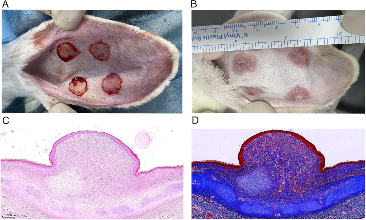
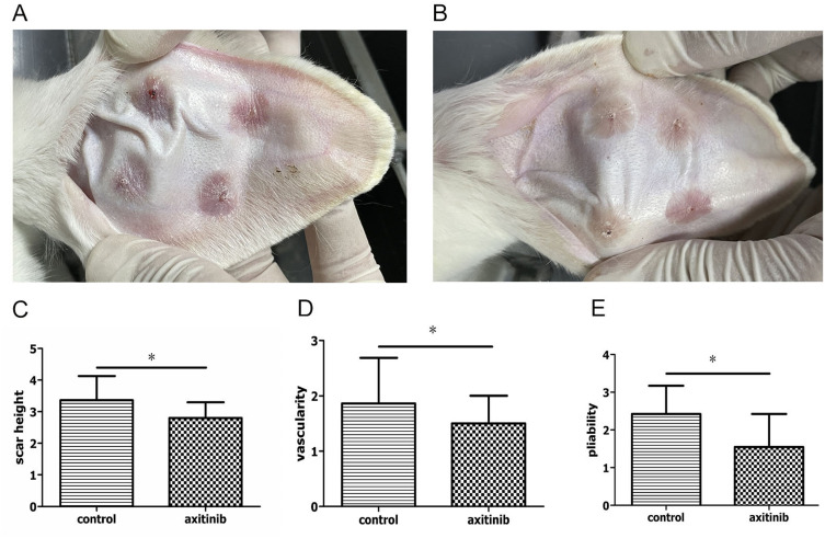
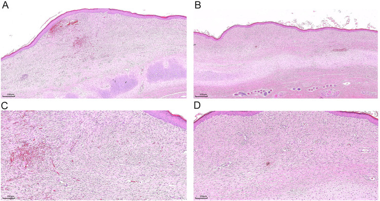
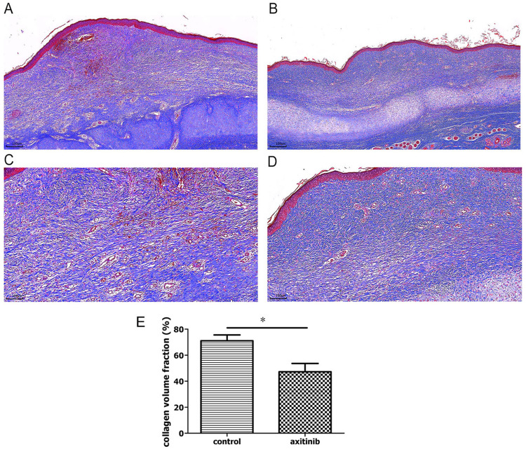
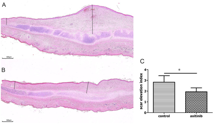
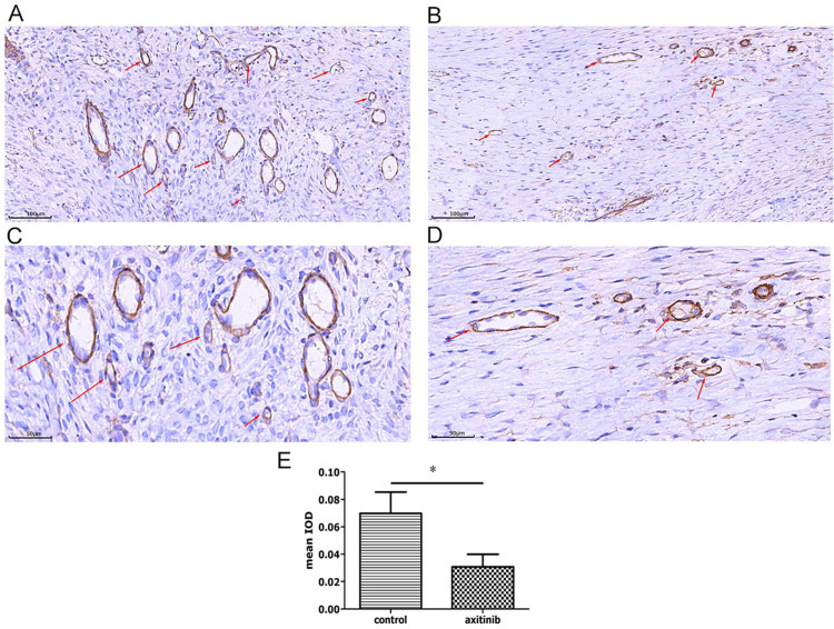
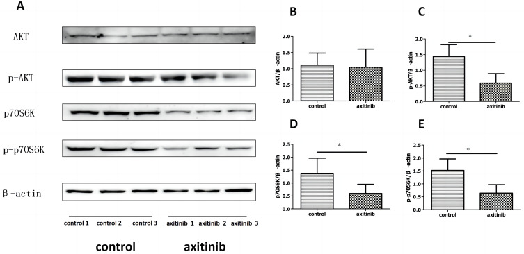
Methods: Ten male New Zealand white rabbits were used to establish HS models and then randomised to the control and axitinib groups. The scar tissues in the two groups were injected with axitinib or normal saline, and they were evaluated after one month of treatment. Macroscopic scar thickness, vascularity and pliability, as well as histopathological analysis including HE staining and Masson staining and scar elevation index (SEI) between two groups were compared. Immunohistochemical staining of CD31 in two groups was conducted to assess the degree of angiogenesis in HS tissue. The protein expression of protein kinase B (AKT) and ribosomal protein S6 kinase (p70S6K) and their phosphorylation levels in both groups were examined by Western blot analysis.

Results: The macroscopic and histological observation showed intralesional axitinib injection significantly reduced scar thickness, vascularity and pliability of HS in the rabbit ear model. The value of SEI in HE assessment was also significantly declined in the axitinib group. Furthermore, immunohistochemical analysis revealed that axitinib suppressed the expression of CD31 in HS tissue, and the mean IOD for blood vessels was significantly lower in the axitinib-treated group. Additionally, axitinib effectively attenuated the protein expression of p70S6K, p-AKT and p-p70S6K by Western blot analysis.

Conclusion: Our study suggests that intralesional injection of axitinib can effectively attenuate HS by reducing angiogenesis in the rabbit ear model, and this inhibitory effect may be mediated by suppression of AKT/p70S6K signaling pathway. It indicates that axitinib may be a promising option for the treatment of HS in the future.

Keywords: angiogenesis; axitinib; hypertrophic scar; rabbit ear scar model.


Conflict of interest statement

The authors report no conflicts of interest in this work.

Figures

Figure 1
Figure 1
Hypertrophic scar (HS) was established in a rabbit ear. (A) Four 15 mm-diameter circular wounds were created on the ventral surface of each ear by removal of tissue deep to the perichondrium. (B) The gross view of HS in the rabbit ear was observed at 30th day after operation. (C and D) The histopathological appearance of HS was shown by HE staining (×25) and Masson staining(×25) respectively.
Figure 2
Figure 2
The inhibitory effect of axitinib on HS in a rabbit ear was observed from gross view. (A and B) The scar tissue was less conspicuous in axitinib group (B) than that in control group (A). (CE) In comparison with HS in the control group, scar thickness (C), vascularity (D) and pliability (E) of HS was significantly decreased in the axitinib group (n = 40 per group). *P < 0.05.
Figure 3
Figure 3
Histopathological images of HE stained HS after treatment. Compared to the control group ((A), ×40, (C), ×100, n = 20), the axitinib group showed decreased and relatively less organised collagen, and reduced fibroblasts and microvessels ((B), ×40, (D), ×100, n = 20).
Figure 4
Figure 4
Histopathological images of Masson stained HS after treatment. (AD) In contrast with that in the control group ((A), ×40, (C), ×100, n = 20), collagen fibers were loose and regularly arranged in the axitinib group ((B), ×40, (D), ×100, n = 20). (E) The value of collagen volume fraction (CVF, %) was significantly decreased in the axitinib group (n = 20 per group). *P < 0.05.
Figure 5
Figure 5
Scar elevation index (SEI) in HE analysis of HS after treatment. (A and B) SEI was calculated in HE staining (×25) between two groups by the ratio of the maximum height of scar to that of surrounding normal skin. (C)The value of SEI was significantly decreased in the axitinib group (n = 20 per group). *P < 0.05.
Figure 6
Figure 6
Axitinib inhibited HS angiogenesis. (AD) Expression of CD31 (as shown by red arrows) in immunohistochemical staining of scar tissues from two groups. CD31 was less in the axitinib group ((B), ×200, (D), ×400) than that in the control group ((A), ×200, (C), ×400). (E) Mean IOD for blood vessels between two groups analyzed by Image-Pro Plus 6. The difference in mean IOD between two groups was significant (n = 20 per group, P = 0.010). *P < 0.05.
Figure 7
Figure 7
Axitinib significantly reduced levels of p-AKT, p70S6K and p-p70S6K shown in Western blot results. (A) The protein levels of AKT and p-AKT, p70S6K and p-p70S6K were determined by immunoblot (n = 6 per group). (B) No significant difference in the relative levels of AKT was detected (P > 0.05). (CE) The relative levels of p-AKT (C), p70S6K (D) and p-p70S6K (E) in the axitinib group were significantly lower than that in the control group (P < 0.05). *P < 0.05.

References

    1. Lee HJ, Jang YJ. Recent understandings of biology, prophylaxis and treatment strategies for hypertrophic scars and keloids. Int J Mol Sci. 2018;19(3):1. doi: 10.3390/ijms19030711 - DOI - PMC - PubMed
    1. Hsu KC, Luan CW, Tsai YW. Review of silicone gel sheeting and silicone gel for the prevention of hypertrophic scars and keloids. Wounds. 2017;29(5):154–158. - PubMed
    1. Seago M, Shumaker PR, Spring LK, et al. Laser treatment of traumatic scars and contractures: 2020 international consensus recommendations. Lasers Surg Med. 2020;52(2):96–116doi: 10.1002/lsm.23201 - DOI - PubMed
    1. Lv K, Liu H, Xu H, et al. Ablative fractional CO(2) laser surgery improving sleep quality, pain and pruritus in adult hypertrophic scar patients: a prospective cohort study. Burns Trauma. 2021;9:tkab023. doi: 10.1093/burnst/tkab023 - DOI - PMC - PubMed
    1. Ledon JA, Savas J, Franca K, Chacon A, Nouri K. Intralesional treatment for keloids and hypertrophic scars: a review. Dermatol Surg. 2013;39(12):1745–1757. doi: 10.1111/dsu.12346 - DOI - PubMed