Review
. 2023 Sep 27;15(9):1841-1857.
doi: 10.4240/wjgs.v15.i9.1841.

Indocyanine green dye and its application in gastrointestinal surgery: The future is bright green

Affiliations
Review

Indocyanine green dye and its application in gastrointestinal surgery: The future is bright green

Zavier Yongxuan Lim et al. World J Gastrointest Surg. .

Abstract

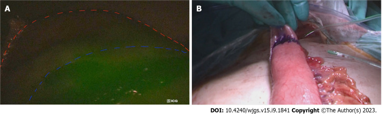
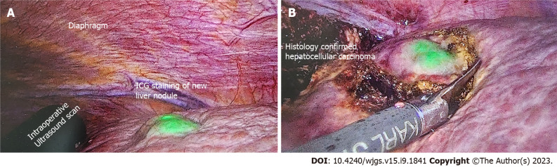
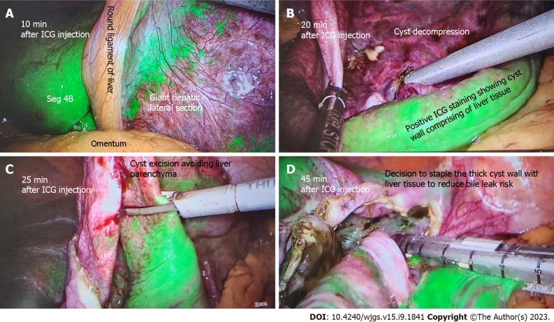
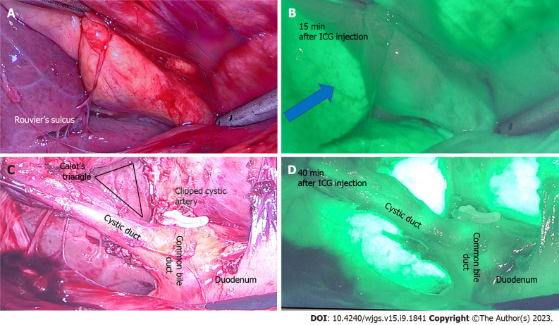
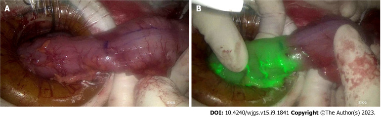
Indocyanine green (ICG) is a water-soluble fluorescent dye that is minimally toxic and widely used in gastrointestinal surgery. ICG facilitates anatomical identification of structures (e.g., ureters), assessment of lymph nodes, biliary mapping, organ perfusion and anastomosis assessment, and aids in determining the adequacy of oncological margins. In addition, ICG can be conjugated to artificially created antibodies for tumour markers, such as carcinoembryonic antigen for colorectal, breast, lung, and gastric cancer, prostate-specific antigen for prostate cancer, and cancer antigen 125 for ovarian cancer. Although ICG has shown promising results, the optimization of patient factors, dye factors, equipment, and the method of assessing fluorescence intensity could further enhance its utility. This review summarizes the clinical application of ICG in gastrointestinal surgery and discusses the emergence of novel dyes such as ZW-800 and VM678 that have demonstrated appropriate pharmacokinetic properties and improved target-to-background ratios in animal studies. With the emergence of robotic technology and the increasing reporting of ICG utility, a comprehensive review of clinical application of ICG in gastrointestinal surgery is timely and this review serves that aim.

Keywords: Fluorescence imaging; Gastrointestinal surgery; Indocyanine green.


Conflict of interest statement

Conflict-of-interest statement: All the authors report no relevant conflicts of interest for this article.

Figures

Figure 1
Figure 1
The utility of indocyanine green dye in oesopgago-gastric anastomosis planning. A: The line of demarcation of indocyanine green (ICG (blue line) at the tip of gastric conduit (red line) to assess perfusion in a patient; B: The prepared gastric conduit with the tip of conduit with poor blood supply, as determined by ICG marked with blue marking line.
Figure 2
Figure 2
A 84-year-old patient with imaging showing 7 cm hepatocellular carcinoma was scheduled for elective laparoscopic right posterior sectionectomy. Indocyanine green dye was injected 10 d before the surgery date. A: Cirrhotic liver with a new liver lesion detected by positive indocyanine green (ICG) staining; B: Excision of this nodule with adequate margins as guided by ICG. Postoperative histology confirmed the new nodule to be primary hepatocellular carcinoma. ICG: Indocyanine green.
Figure 3
Figure 3
A 60-year-old patient undergoing elective liver cyst deroofing for a symptomatic solitary benign epithelial liver cyst was injected with 7 mL of indocyanine green dye after insertion of camera port. A: Liver enhancement at 10 min; B: After 20 min of injection shows the dye enhances the liver and cyst wall remains unenhanced; C: How indocyanine green (ICG) guidance can avoid transecting the liver parenchyma during cyst wall excision; D: The cyst wall with positive ICG staining is excised using stapling technology to reduce bile leak risk. ICG: Indocyanine green.
Figure 4
Figure 4
A 50-year-old patient undergoing elective laparoscopic cholecystectomy for previous acute cholecystitis was injected with 4 mL of indocyanine green dye after insertion of camera port. A: Rouvier’s sulcus and corresponding; B: After 15 min of injection shows the dye enhances the liver (blue arrow) and indocyanine green (ICG) is yet to be excreted in biliary tree; C: Calot’s triangle with a critical view of safety and clipped cystic artery; D: At 40 min after ICG injection shows beginning of biliary excretion in cystic duct and common bile duct. ICG: Indocyanine green.
Figure 5
Figure 5
The utility of indocyanine green dye in laparoscopic anterior resection. A: The descending colon prepared for proximal transection during laparoscopic anterior resection, with the purple line indicating intended transection site, 5 cm proximal to tumour; B: The indocyanine green angiography confirms good vascularity at the site of intended transection, prior to creation of colo-rectal anastomosis.

References

    1. Lu CH, Hsiao JK. Indocyanine green: An old drug with novel applications. Tzu Chi Med J. 2021;33:317–322. - PMC - PubMed
    1. Reinhart MB, Huntington CR, Blair LJ, Heniford BT, Augenstein VA. Indocyanine Green: Historical Context, Current Applications, and Future Considerations. Surg Innov. 2016;23:166–175. - PubMed
    1. Kraft JC, Ho RJ. Interactions of indocyanine green and lipid in enhancing near-infrared fluorescence properties: the basis for near-infrared imaging in vivo. Biochemistry. 2014;53:1275–1283. - PMC - PubMed
    1. Lund-Johansen P. The dye dilution method for measurement of cardiac output. Eur Heart J. 1990;11 Suppl I:6–12. - PubMed
    1. Destro M, Puliafito CA. Indocyanine green videoangiography of choroidal neovascularization. Ophthalmology. 1989;96:846–853. - PubMed