Lower-limb internal loading and potential consequences for fracture healing
- PMID: 37901836
- PMCID: PMC10602681
- DOI: 10.3389/fbioe.2023.1284091
Lower-limb internal loading and potential consequences for fracture healing
Abstract
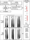
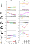
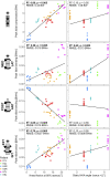
Introduction: Mechanical loading is known to determine the course of bone fracture healing. We hypothesise that lower limb long bone loading differs with knee flexion angle during walking and frontal knee alignment, which affects fracture healing success. Materials and methods: Using our musculoskeletal in silico modelling constrained against in vivo data from patients with instrumented knee implants allowed us to assess internal loads in femur and tibia. These internal forces were associated with the clinical outcome of fracture healing in a relevant cohort of 178 extra-articular femur and tibia fractures in patients using a retrospective approach. Results: Mean peak forces differed with femoral compression (1,330-1,936 N at mid-shaft) amounting to about half of tibial compression (2,299-5,224 N). Mean peak bending moments in the frontal plane were greater in the femur (71-130 Nm) than in the tibia (from 26 to 43 Nm), each increasing proximally. Bending in the sagittal plane showed smaller mean peak bending moments in the femur (-38 to 43 Nm) reaching substantially higher values in the tibia (-63 to -175 Nm) with a peak proximally. Peak torsional moments had opposite directions for the femur (-13 to -40 Nm) versus tibia (15-48 Nm) with an increase towards the proximal end in both. Femoral fractures showed significantly lower scores in the modified Radiological Union Scale for Tibia (mRUST) at last follow-up (p < 0.001) compared to tibial fractures. Specifically, compression (r = 0.304), sagittal bending (r = 0.259), and frontal bending (r = -0.318) showed strong associations (p < 0.001) to mRUST at last follow-up. This was not the case for age, body weight, or localisation alone. Discussion: This study showed that moments in femur and tibia tend to decrease towards their distal ends. Tibial load components were influenced by knee flexion angle, especially at push-off, while static frontal alignment played a smaller role. Our results indicate that femur and tibia are loaded differently and thus require adapted fracture fixation considering load components rather than just overall load level.
Keywords: femur; fracture fixation; in vivo loading; internal bone loading; intramedullary nail; locking plate; musculoskeletal modelling; tibia.
Copyright © 2023 Heyland, Deppe, Reisener, Damm, Taylor, Reinke, Duda and Trepczynski.
Conflict of interest statement
MH reports grants from Stryker, during the conduct of the study. PD reports grants from the OrthoLoadClub, during the conduct of the study. GD reports grants from Pluristem, DePuy Synthes, Implantec, Implantcast, S&N, Stryker, Zimmer, outside the submitted work. The remaining authors declare that the research was conducted in the absence of any commercial or financial relationships that could be construed as a potential conflict of interest.
Figures


References
LinkOut - more resources
Full Text Sources

