. 2024 Jun;39(3):113-124.
doi: 10.1111/omi.12442. Epub 2023 Oct 30.

Primed inflammatory response by fibroblast subset is necessary for proper oral and cutaneous wound healing

Affiliations

Primed inflammatory response by fibroblast subset is necessary for proper oral and cutaneous wound healing

Zhaoxu Chen et al. Mol Oral Microbiol. 2024 Jun.

Abstract

Fibroblasts are ubiquitous mesenchymal cells that exhibit considerable molecular and functional heterogeneity. Besides maintaining stromal integrity, oral fibroblast subsets are thought to play an important role in host-microbe interaction during injury repair, which is not well explored in vivo. Here, we characterize a subset of fibroblast lineage labeled by paired-related homeobox-1 promoter activity (Prx1Cre+) in oral mucosa and skin and demonstrate these fibroblasts readily respond to microbial products to facilitate the normal wound healing process. Using a reporter mouse model, we determined that Prx1Cre+ fibroblasts had significantly higher expression of toll-like receptors 2 and 4 compared to other fibroblast populations. In addition, Prx1 immunopositive cells exhibited heightened activation of inflammatory transcription factor NF-κB during the early wound healing process. At the cytokine level, CXCL1 and CCL2 were significantly upregulated by Prx1Cre+ fibroblasts at baseline and upon LPS stimulation. Importantly, lineage-specific knockout to prevent NF-κB activation in Prx1Cre+ fibroblasts drastically impaired both oral and skin wound healing processes, which was linked to reduced macrophage infiltration, failure to resolve inflammation, and clearance of bacteria. Together, our data implicate a pro-healing role of Prx1-lineage fibroblasts by facilitating early macrophage recruitment and bacterial clearance.

Keywords: Prx1; fibroblast; immunomodulation; inflammation; regeneration; wound healing.


Conflict of interest statement

Conflict of Interest

The authors have no conflict of interest to declare.

Figures
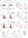
Figure 1.
Figure 1.
Prx1Cre+ fibroblasts highly express TLR2 and TLR4. A. Flow cytometry plot gated for fibroblasts (live, CD31, CD45, EpCam, Ter119, PDGFRA+) to identify Prx1Cre+ and Prx1Cre lineage cells by their co-expression of tdTomato (tdTom) in Prx1Cre+:R26RtdTomato mice. B. Quantification of tdTom+ Prx1Cre+ and tdTom Prx1Cre fibroblast populations from palatal gingiva and scalp skin. N = 3 mice each. C. Left, representative flow cytometry mean fluorescence intensity (MFI) histogram for TLR2 and TLR4 expression in oral Prx1Cre+ and Prx1Cre fibroblasts from unwounded gingiva in Prx1Cre+:R26RtdTomato mice. Right, quantification of TLR2 and TLR4 MFI in oral Prx1Cre+ and Prx1Cre fibroblasts. N = 4 mice each. D. Left, representative flow cytometry MFI histogram for TLR2 and TLR4 expression in skin Prx1Cre+ and Prx1Cre fibroblasts from unwounded scalp in Prx1Cre+:R26RtdTomato mice. Right, quantification of TLR2 and TLR4 MFI in skin Prx1Cre+ and Prx1Cre fibroblasts. N = 4 mice per group. E. Quantification of Prx1 protein expression in TLR2, TLR4, PDGFRA+ fibroblasts (black), TLR2+ PDGFRA+ fibroblasts (light red), and TLR4+ PDGFRA+ fibroblasts (red) in oral mucosa (left) and scalp skin (right) of wildtype B6 mice. N = 3 mice each. A.U.: arbitrary unit. Student’s t-test, *P < 0.05, **P < 0.01.
Figure 2.
Figure 2.
Enhanced nuclear NF-κB expression in Prx1Cre+ fibroblasts under wounded conditions. A. Representative immunofluorescence image of day 4 oral wound stained with vimentin, NF-κB (p65) and Prx1 antibodies. Scale bar, 100 μm. Inset shows an area adjacent to original oral wound, scale bar, 50 μm. B. Quantification of nuclear NF-κB+ fibroblasts that are either Prx1+ or Prx1 in day 4 oral wounds. C. Representative immunofluorescence image of day 4 scalp skin wound stained with vimentin, NF-κB and Prx1 antibodies. Scale bar, 100 μm. Inset shows an area adjacent to original skin wound, scale bar, 50 μm. D. Quantification of nuclear NF-κB+ fibroblasts that are either Prx1+ or Prx1 in day 4 skin wounds. Filled arrowheads point to nuclear NF-κB expression in Prx1+ fibroblasts (DAPI+ nuclear NF-κB+ Prx1+ vimentin+ spindle-shaped cells), and empty arrowheads point to nuclear NF-κB expression in Prx1- fibroblasts (DAPI+ nuclear NF-κB+ Prx1 vimentin+ spindle-shaped cells). N = 3 mice. Student’s t-test, **P < 0.01, ***P < 0.001.
Figure 3.
Figure 3.
Cytokine expression profile in Prx1Cre+ fibroblasts. A. Experimental scheme for sorting Prx1Cre+ and Prx1Cre fibroblasts from palatal gingiva of Prx1Cre+:R26RtdTomato mice. B. RT-qPCR of Cxcl1, Ccl2, Il6, and Il1b comparing sorted Prx1Cre+ fibroblasts and Prx1Cre fibroblasts, normalized to L32 expression. N = 3 mice. Student’s t-test, ****P < 0.0001. C. Representative mean fluorescence intensity (MFI) plot of CXCL1, CCL2, IL6, and IL1β expression in Prx1Cre+ and Prx1Cre cells after 6 hrs of LPS (5ug/ml) stimulation. D. Quantification of CXCL1, CCL2, IL6, and IL1β expression by Prx1Cre+ and Prx1Cre cells after 6 hrs of LPS (5ug/ml) stimulation in vitro. Data represent mean ± SEM. Experiments were performed in triplicate (N = 3) and in two independent experiments with similar results. One-way ANOVA, *P < 0.05.
Figure 4.
Figure 4.
Lineage specific deletion of NF-κB activator kinase Ikkβ delays wound healing. A. Representative microphotographs of 1-mm oral wound 7 days post wounding in either Ikkβf/f (control) or Prx1Cre+:Ikkβf/f (experimental) mice. Scale bar, 1 mm. B. Quantification of clinically open oral wound area 7 days post wounding in mm2. C. H&E-stained sections of day 7 oral wound in control and experimental mice. Arrow designates original 1-mm wound length. Scale bar, 0.5 mm. D. Quantification of epithelial gap of oral wound 7 days post wounding in mm. E. Left, trichrome stained sections of day 7 oral wound in control and experimental mice. Arrowheads point to the wound edge. Dashed lines demarcates new connective tissue area. Scale bar, 50 μm. Right, quantification of new oral connective tissue area in mm2. F. Representative microphotographs of 2-mm scalp wound 7 days post wounding in control and experimental mice. Scale bar, 2 mm. G. Quantification of clinically open skin wound area 7 days post wounding in mm2. H. H&E-stained sections of day 7 scalp skin wound in control and experimental mice. Arrow designates original 2-mm wound length. Scale bar, 1 mm. I. Quantification of epithelial gap of skin wound 7 days post wounding in mm. J. Left, trichrome stained sections of day 7 scalp wound in control and experimental mice. Arrowheads point to the wound edge. Dashed lines demarcates new dermal connective tissue. Scale bar, 100 μm. Right, quantification of new dermal connective tissue area in mm2. A-J, N = 5–7 per group for a total of 12 mice. Student’s t-test, *P < 0.05, **P < 0.01, ***P < 0.001.
Figure 5.
Figure 5.
Perturbation of proinflammatory function in Prx1Cre+ fibroblasts prevent resolution of inflammation and bacterial clearance. A. Left, representative immunofluorescence image of day 7 gingival wounds in Ikkβf/f (control) or Prx1Cre+:Ikkβf/f (experimental) mice stained with antibodies against CD45 and K14. Right, quantification of CD45+ leukocytes normalized to total nucleated cell number in lamina propria. B. Left, representative RNAscope image of day 7 gingival wounds in control and experimental mice hybridized with eubacterial 16S rRNA probe. Right, quantification of RNA puncta normalized to total nucleated cell number in lamina propria. C. Left, representative immunofluorescence image of day 7 skin wounds in control and experimental mice stained with antibodies against CD45 and K14. Right, quantification of CD45+ leukocytes normalized to total nucleated cell number in skin dermis. D. Left, representative RNAscope image of day 7 skin wounds in control and experimental mice hybridized with eubacterial 16S rRNA probe. Right, quantification of RNA puncta normalized to total nucleated cell number in skin dermis. E. Representative flow cytometry plot pre-gated for CD45+ live cells from day 4 oral wounds, and F. Quantification of percent Ly6g+ neutrophils (left) and F4/80+ macrophages (right) normalized to CD45+ cell numbers from day 4 oral wounds of control and experimental mice. PMN, polymorphonuclear neutrophils, MΦ, macrophages. G. Representative flow cytometry plot pre-gated for CD45+ live cells from day 4 skin wounds, and H. Quantification of percent Ly6g+ neutrophils (left) and F4/80+ macrophages (right) normalized to CD45+ cell numbers from day 4 skin wounds of control and experimental mice. A-D, Dashed lines demarcates epithelium-dermis boarder, scale bar 50 μm. N = 5–7 per group for a total of 12 mice. E-H, N = 4–5 per group for a total of 9–10 mice. Student’s t-test, *P < 0.05, **P < 0.01, ***P < 0.001.

Similar articles

Cited by

References

    1. Bassir SH, Garakani S, Wilk K, Aldawood ZA, Hou J, Yeh SA, Sfeir C, Lin CP, & Intini G (2019). Prx1 Expressing Cells Are Required for Periodontal Regeneration of the Mouse Incisor. Front Physiol, 10, 591. 10.3389/fphys.2019.00591 - DOI - PMC - PubMed
    1. Boothby IC, Kinet MJ, Boda DP, Kwan EY, Clancy S, Cohen JN, Habrylo I, Lowe MM, Pauli M, Yates AE, Chan JD, Harris HW, Neuhaus IM, McCalmont TH, Molofsky AB, & Rosenblum MD (2021). Early-life inflammation primes a T helper 2 cell-fibroblast niche in skin. Nature. 10.1038/s41586-021-04044-7 - DOI - PMC - PubMed
    1. Brinson CW, Lu Z, Li Y, Lopes-Virella MF, & Huang Y (2016). Lipopolysaccharide and IL-1beta coordinate a synergy on cytokine production by upregulating MyD88 expression in human gingival fibroblasts. Mol Immunol, 79, 47–54. 10.1016/j.molimm.2016.09.020 - DOI - PMC - PubMed
    1. Buechler MB, Pradhan RN, Krishnamurty AT, Cox C, Calviello AK, Wang AW, Yang YA, Tam L, Caothien R, Roose-Girma M, Modrusan Z, Arron JR, Bourgon R, Muller S, & Turley SJ (2021). Cross-tissue organization of the fibroblast lineage. Nature, 593(7860), 575–579. 10.1038/s41586-021-03549-5 - DOI - PubMed
    1. Croft AP, Campos J, Jansen K, Turner JD, Marshall J, Attar M, Savary L, Wehmeyer C, Naylor AJ, Kemble S, Begum J, Durholz K, Perlman H, Barone F, McGettrick HM, Fearon DT, Wei K, Raychaudhuri S, Korsunsky I, Brenner MB, Coles M, Sansom SN, Filer A, & Buckley CD (2019). Distinct fibroblast subsets drive inflammation and damage in arthritis. Nature, 570(7760), 246–251. 10.1038/s41586-019-1263-7 - DOI - PMC - PubMed

MeSH terms

LinkOut - more resources