. 2023 Nov 7;120(45):e2312022120.
doi: 10.1073/pnas.2312022120. Epub 2023 Oct 30.

Lateral interactions govern self-assembly of the bacterial biofilm matrix protein BslA

Affiliations

Lateral interactions govern self-assembly of the bacterial biofilm matrix protein BslA

Sofia Arnaouteli et al. Proc Natl Acad Sci U S A. .

Abstract

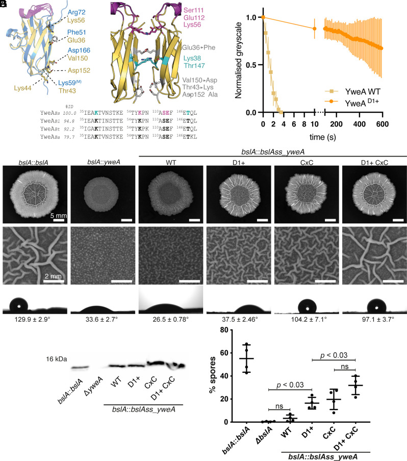
The soil bacterium Bacillus subtilis is a model organism to investigate the formation of biofilms, the predominant form of microbial life. The secreted protein BslA self-assembles at the surface of the biofilm to give the B. subtilis biofilm its characteristic hydrophobicity. To understand the mechanism of BslA self-assembly at interfaces, here we built a molecular model based on the previous BslA crystal structure and the crystal structure of the BslA paralogue YweA that we determined. Our analysis revealed two conserved protein-protein interaction interfaces supporting BslA self-assembly into an infinite 2-dimensional lattice that fits previously determined transmission microscopy images. Molecular dynamics simulations and in vitro protein assays further support our model of BslA elastic film formation, while mutagenesis experiments highlight the importance of the identified interactions for biofilm structure. Based on this knowledge, YweA was engineered to form more stable elastic films and rescue biofilm structure in bslA deficient strains. These findings shed light on protein film assembly and will inform the development of BslA technologies which range from surface coatings to emulsions in fast-moving consumer goods.

Keywords: Bacillus subtilis; X-ray crystallography; biofilm matrix; molecular dynamic simulations; protein assemblies.


Conflict of interest statement

The authors disclose the following patent filing: US20170267730A1.

Figures

Fig. 1.
Fig. 1.
YweA adopts a β-sandwich fold similar to BslA. (A) Cartoon representation of YweA (PDB 5MKD, chain B) colored by secondary structure. The β-strands are yellow, α-helices teal, and loop regions in gray. The N- and C-termini are labeled (“N” and “C” respectively) along with loops (L), β-strands (β), and α-helices (α), which are numbered by when they occur within the primary amino acid sequence. (B) Structural alignment of the four monomers of the asymmetric unit from the side of the sandwich and from the top showing the variation in loops 2, 3, 4, and 6 between monomers. Sidechains of the poorest aligning residues are displayed (chain A: light gray, B: yellow, C: medium gray, D: dark gray). (C) Superposition of BslA (PDB 4BHU, chain C, blue) onto YweA (PDB 5MKD, chain B, yellow) shows the structural similarity between the core Ig-fold of the two paralogues. (D) Comparison of loops L2, L4, and L6, of YweA to the cap region of BslA shows similar exposed hydrophobic residues but differences in secondary structure. The cap residues are colored purple, whereas the other secondary structure elements are colored yellow for YweA and blue for BslA. Alignment of the three cap regions of YweA with BslA shows conservation of hydrophobic residues. Cap regions are numbered based on their location in the primary amino acid sequence (Cap1 to 3). Sequence identity (%ID) is listed as compared to YweA from B. subtilis (YweABs) calculated over the full sequence alignment. Sequences of YweA homologues from Bacillus licheniformis (Bl), Bacillus tequilensis (Bt), and Bacillus amyloliquefaciens (Ba) as well as BslA from B. subtilis (blue) were included. The amino acid number is listed for the first residue in each cap region. (E). Surface representation of YweA showing surface hydrophobicity colored from least hydrophobic (white) to most hydrophobic (red) based on the Eisenburg hydrophobicity scale.
Fig. 2.
Fig. 2.
The crystal structures of BslA and YweA cap-aligned dimers. (A) Cartoon representation of the BslA asymmetric unit that consists of 10 monomers. Two dimers are present that have cap regions (purple) aligned with their partners (H with C [blue] and G with D [light gray]). (B) Cartoon representation of dimer1 (chain C and H) of BslA PDB 4BHU. BslA is displayed in blue with the cap region in purple. Residues that promote interaction at the interface are displayed with sidechains showing the stacking of Arg72 (yellow), π–π stacking of Phe51 (orange), and positioning of the methylated Lys59 (Lys59(M)) within hydrogen bonding distance of Asp166 (cyan). (C) Cartoon representation of the YweA asymmetric unit that consists of four monomers. Two dimers are present that have cap regions (purple) aligned with their partners [A with B (yellow) and C with D (light gray)]. (D) Cartoon representation of dimer2 containing chain A and B of YweA PDB 5MKD. YweA is displayed in yellow with the cap region in purple. Residues that interact at the interface are shown in stick representation and colored by interacting groups. The Inset shows this zoomed in with residues labeled.
Fig. 3.
Fig. 3.
Construction of a model BslA lattice. (A) Sequence alignment of BslA homologues (Bs: B. subtilis, Bt: B tequilensis, Bl:B. licheniformis, and Bp: B. pumilus) with YweA from B. subtilis shows conservation of some of the dimer2 interface residues between paralogues. Residues that are shown in panel B are in bold and colored accordingly. Sequence identity is listed based on alignment over the entire sequence as compared with BslABs. The first residue of each region is numbered based on its location in the primary sequence. (B and C) Structural model of BslA dimer2 created from the alignment of BslA monomers onto the YweA dimer2. Cap regions are shown in purple. YweA is in yellow and BslA is in blue. Residues that could interact at the BslA dimer interface are colored and labeled with the YweA residues in gray (panel C only). The amino acid labeling refers to BslA. (D) A model trimer created from aligning a monomer of dimer1 with a monomer of dimer2. The dimer interfaces are shown with dashed lines and labeled. (E) A 2-dimensional BslA lattice was produced based on the crystallographic dimers described herein and the TEM data from Bromley et al. (11) The first step (i) was the fast Fourier transform (FFT) to determine the repeat unit of the ordered BslA lattice (x = 4.3, y = 3.9, α = β = 90°). Step (ii) was the averaging of the TEM image to see four repeat units (octamer) where brighter pixels represent higher electron density. The modeled trimer (dimer1circled in purple and dimer2 circled in green) is congruent with the TEM pattern (iii) A lattice can be constructed (iv) by the structural alignment of monomers from the crystallographic dimers which creates a chain across the page. Translation of the propagated dimers by 3.9 nm (y direction) leads to a hypothetical 2D lattice that could extend infinitely. The repeating units measure 4.3 nm by 3.9 nm in agreement with the FFT. The lattice is shown with the caps (purple) all facing out of the page. Verification of the model (v) was done by comparison of the electron density of the model (converted to hypothetical TEM intensity) with the intensity of the TEM image over each pixel shows a good correlation.
Fig. 4.
Fig. 4.
Molecular dynamic simulations support dimer orientations. (A) Snapshots of the MD simulation at equilibrium (t = 100 ns) for BslA dimer1 (purple) and dimer2 (green) in an aqueous box with an air interface. (B) Orientation of BslA monomer (black), dimer1 (purple circles), and dimer2 (green triangles) in relation to the normal of the interface as determined from equilibrium MD simulations. C. Measure of the distance (nm) between BslA monomers during nonbiased equilibrium MD simulations for the first 35 ns, for Dimer1 (dark purple), Dimer2 (dark green), BslAD1– (light purple), and BslAD2– (light green). (D) PMF is indicative of the binding affinities of the dimer1 interface (purple) and the dimer2 interface (green). The force in kcal/mol was graphed against the change in distance (nm) from the equilibrium position of the pulled monomer. The bars represent the errors on our PMF estimate. (E) wrinkle relaxation graphed by the change in normalized grayscale of pendent drop wrinkles over time (s) for BslA WT (blue), BslAD1– (light purple), and BslAD2– (light green). Error bars represent SD. (F) Representative colony biofilms and sessile drop images. Hydrophobicity measures are listed representing the angle of the edge of the droplet to the base n = 3, errors represent the SEM. A contact angle of greater than 90° is indicative of a nonwetting/hydrophobic surface. (G) Immunoblot of the matrix localized BslA variant from the strains analyzed in F.
Fig. 5.
Fig. 5.
Engineering of YweA dimer1 interface increases film stability. (A) Alignment of YweA (yellow) and BslA (blue) shows that the BslA dimer1 residues (gray, labeled in blue) are not conserved. Cap regions are colored in purple for orientation. Aligned YweA residues and amino acid labels are shown in yellow. (B) The model of the YweA dimer1 interface shows a possible hydrogen bonding network. The residues of note are pink and cyan. Residues in gray are those mutated to create the BslA-like dimer1 interface on YweA. (C) Sequence alignment of YweA homologues (Bs: B. subtilis, Bt: B tequilensis, Bl: B. licheniformis, and Ba: B. amyloliqufaciens) shows conservation of the dimer1 interface residues. Residues that are shown as sticks in panel B are in bold and colored accordingly. Sequence identity is listed based on alignment over the entire sequence as compared with YweABs. The first residue of each region is numbered based on its location in the primary sequence. (D) YweA film relaxation after droplet compression (graphed by the normalized gray value) over time. YweA WT is graphed in yellow (squares) and the YweAD1+ mutant is in orange (circles). (E) Representative colony biofilm images as well as zoomed-in in the center of each colony with hydrophobicity measures listed below. A contact angle of greater than 90° is indicative of a nonwetting/hydrophobic surface. (F) α-YweA immunoblot of matrix proteins from the biofilms including a yweA deficient sample (NRS2405) as a negative control. (G) Quantification of heat-resistant spores (sporulation) for each strain when grown as a colony biofilm plotted as the mean value over four biological replicates. Error bars are the SD and statistics are shown as calculated from a one-way ANOVA with Šídák's multiple comparisons test in GraphPad Prism 9.
Fig. 6.
Fig. 6.
Model of BslA film formation. BslA exists in solution in a soluble “cap in” configuration [for simplicity, BslA is only shown in its monomeric form but in solution can also exist as dimers and tetramers mediated via disulfide bonds through their CxC motifs, unrelated to the interfacial lateral interactions (13)]. BslA absorbs onto an interface and undergoes a limited structural rearrangement into the “cap out” form (11), which exposes the cap hydrophobic residues and reorientates the protein (12), facilitating lateral self-assembly. The final cartoon illustrates the D1 and D2 lateral interactions that our experimental in vitro and in vivo data support as the molecular basis for interactions between monomers that hold the film together.

References

    1. Flemming H. C., Wuertz S., Bacteria and archaea on Earth and their abundance in biofilms. Nat. Rev. Microbiol. 17, 247–260 (2019). - PubMed
    1. Arnaouteli S., Bamford N. C., Stanley-Wall N. R., Kovacs A. T., Bacillus subtilis biofilm formation and social interactions. Nat. Rev. Microbiol. 19, 600–614 (2021). - PubMed
    1. Karygianni L., Ren Z., Koo H., Thurnheer T., Biofilm matrixome: Extracellular components in structured microbial communities. Trends Microbiol. 28, 668–681 (2020). - PubMed
    1. Romero D., Aguilar C., Losick R., Kolter R., Amyloid fibers provide structural integrity to Bacillus subtilis biofilms. Proc. Natl. Acad. Sci. U.S.A. 107, 2230–2234 (2010). - PMC - PubMed
    1. Erskine E., et al. , Formation of functional, non-amyloidogenic fibres by recombinant Bacillus subtilis TasA. Mol. Microbiol. 110, 897–913 (2018). - PMC - PubMed

Publication types

MeSH terms

Substances

LinkOut - more resources