. 2023 Nov 7;120(45):e2306395120.
doi: 10.1073/pnas.2306395120. Epub 2023 Oct 30.

The CRL3gigaxonin ubiquitin ligase-USP15 pathway governs the destruction of neurofilament proteins

Affiliations

The CRL3gigaxonin ubiquitin ligase-USP15 pathway governs the destruction of neurofilament proteins

Hyoung-Min Park et al. Proc Natl Acad Sci U S A. .

Abstract

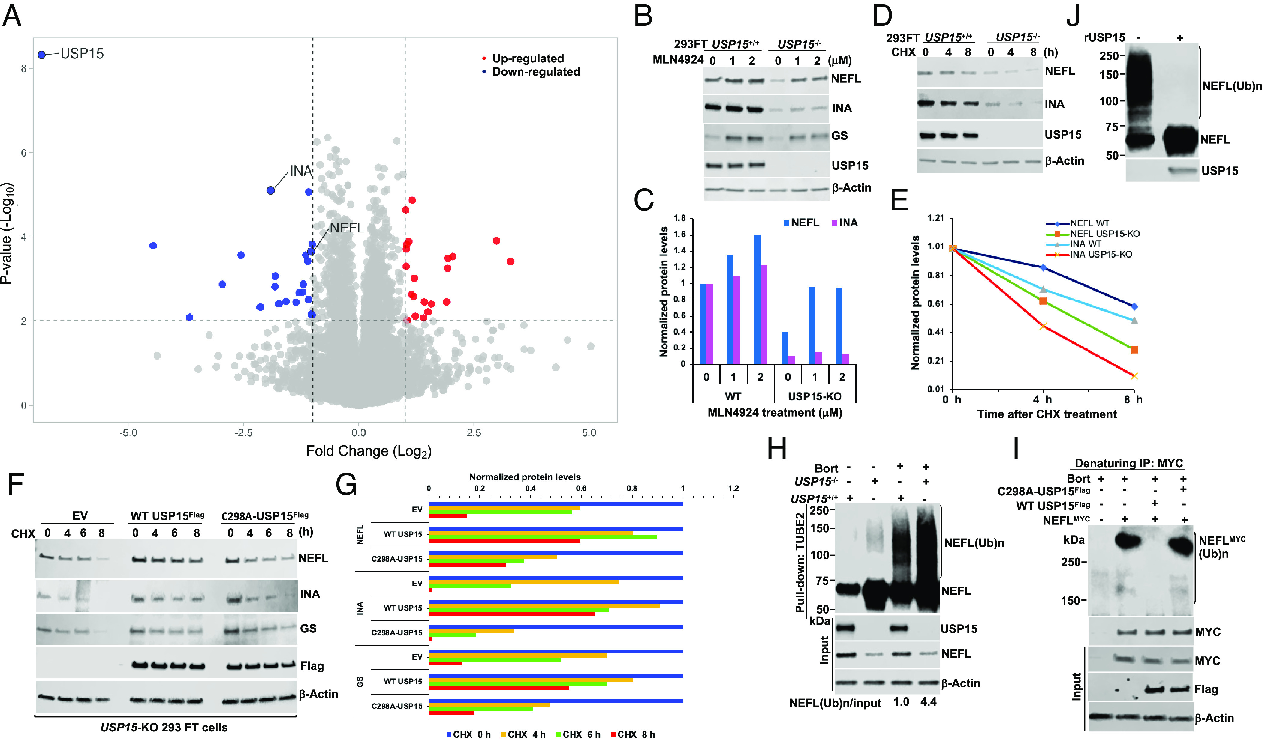
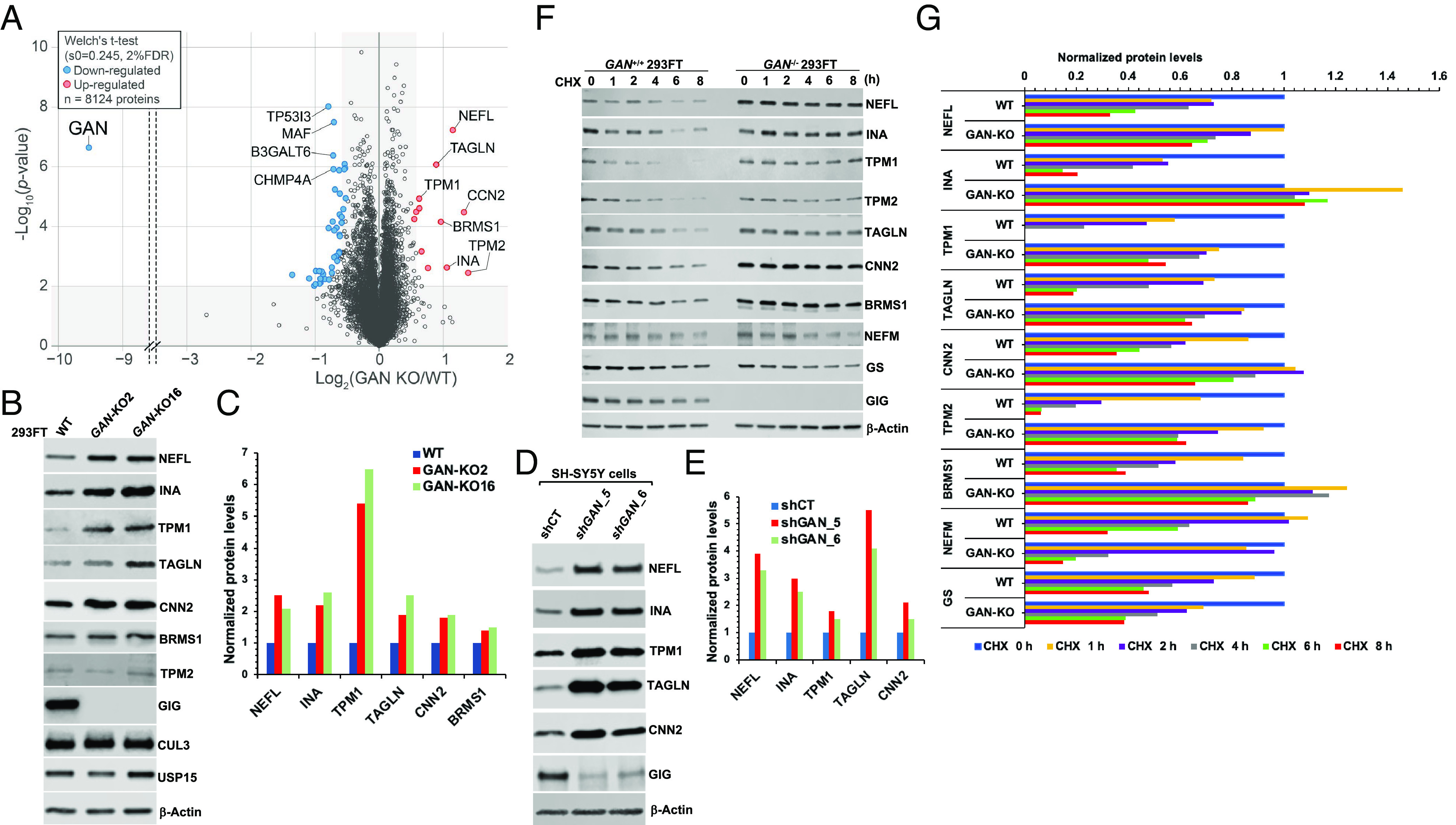
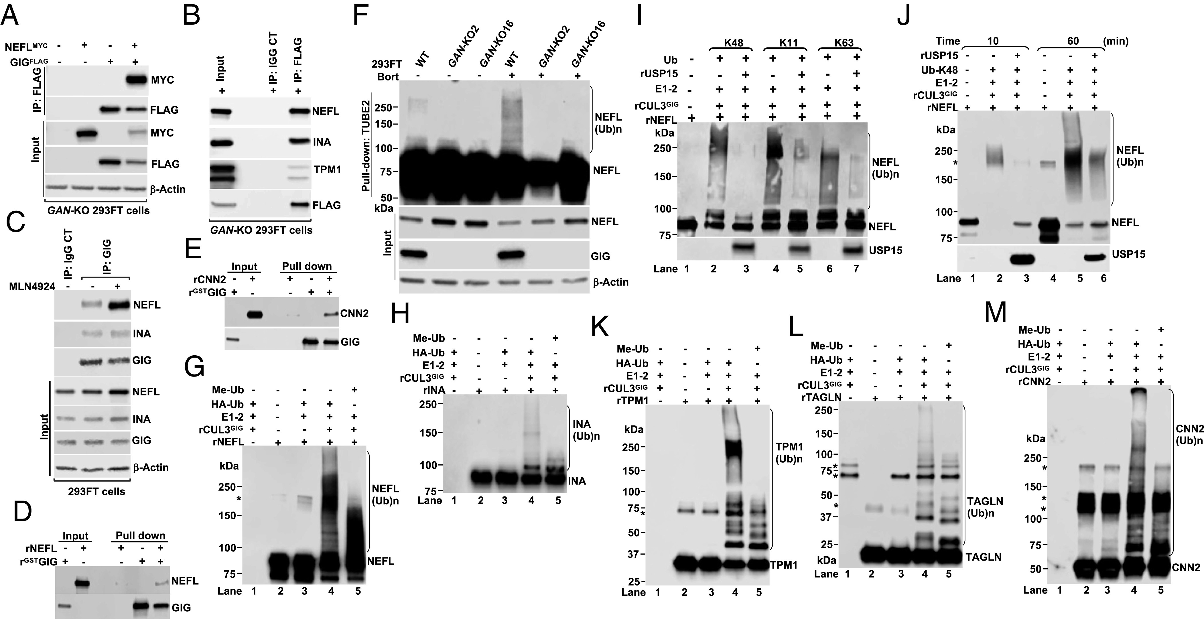
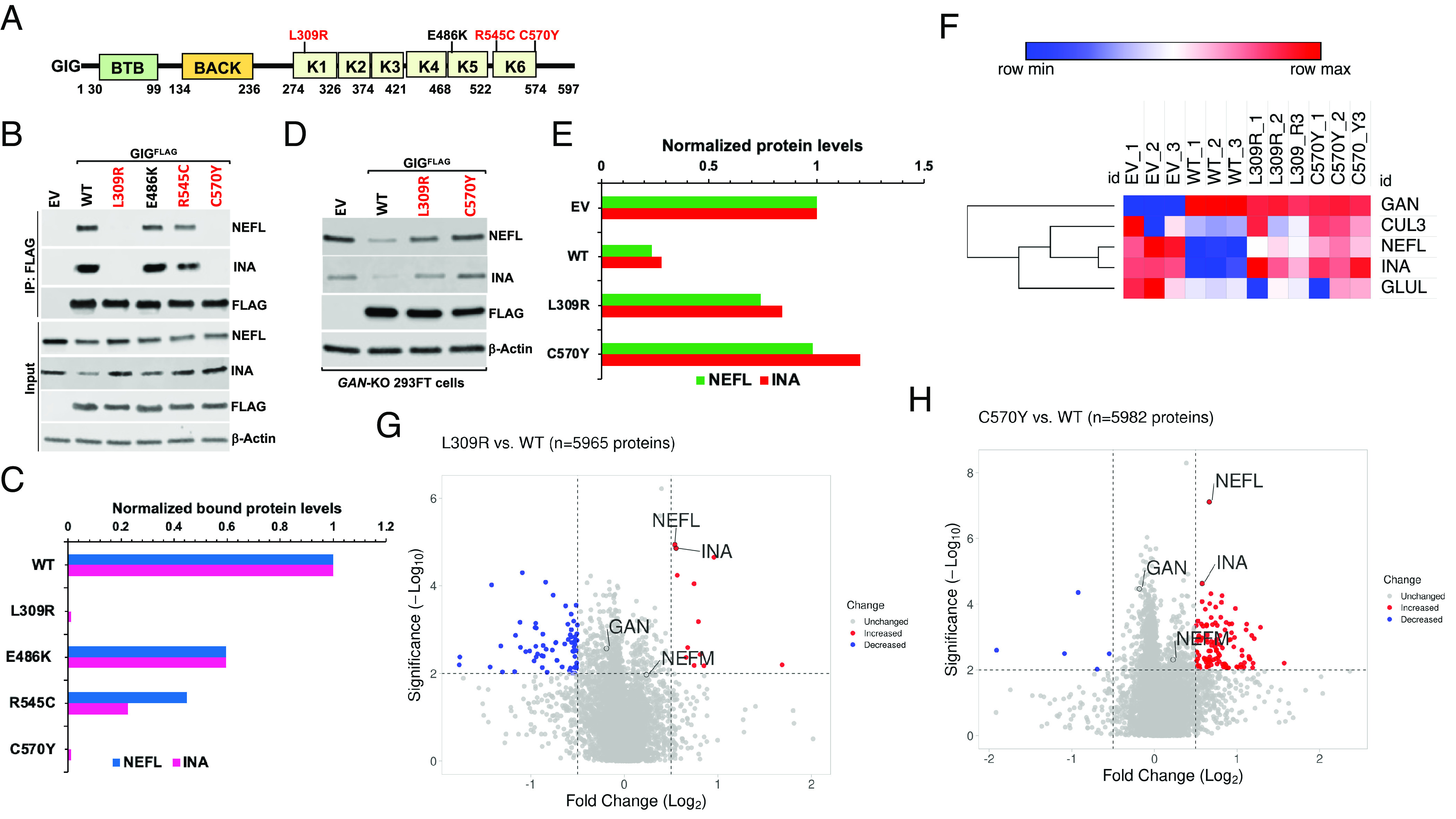
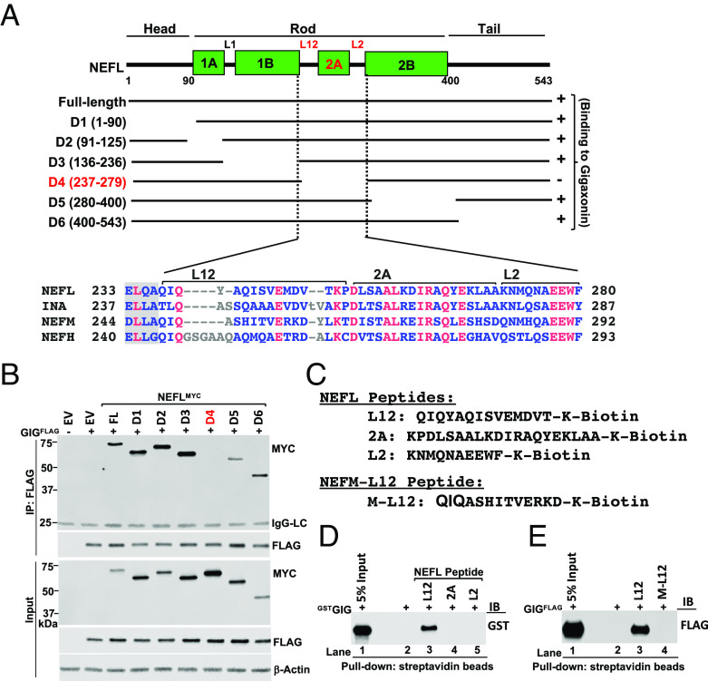
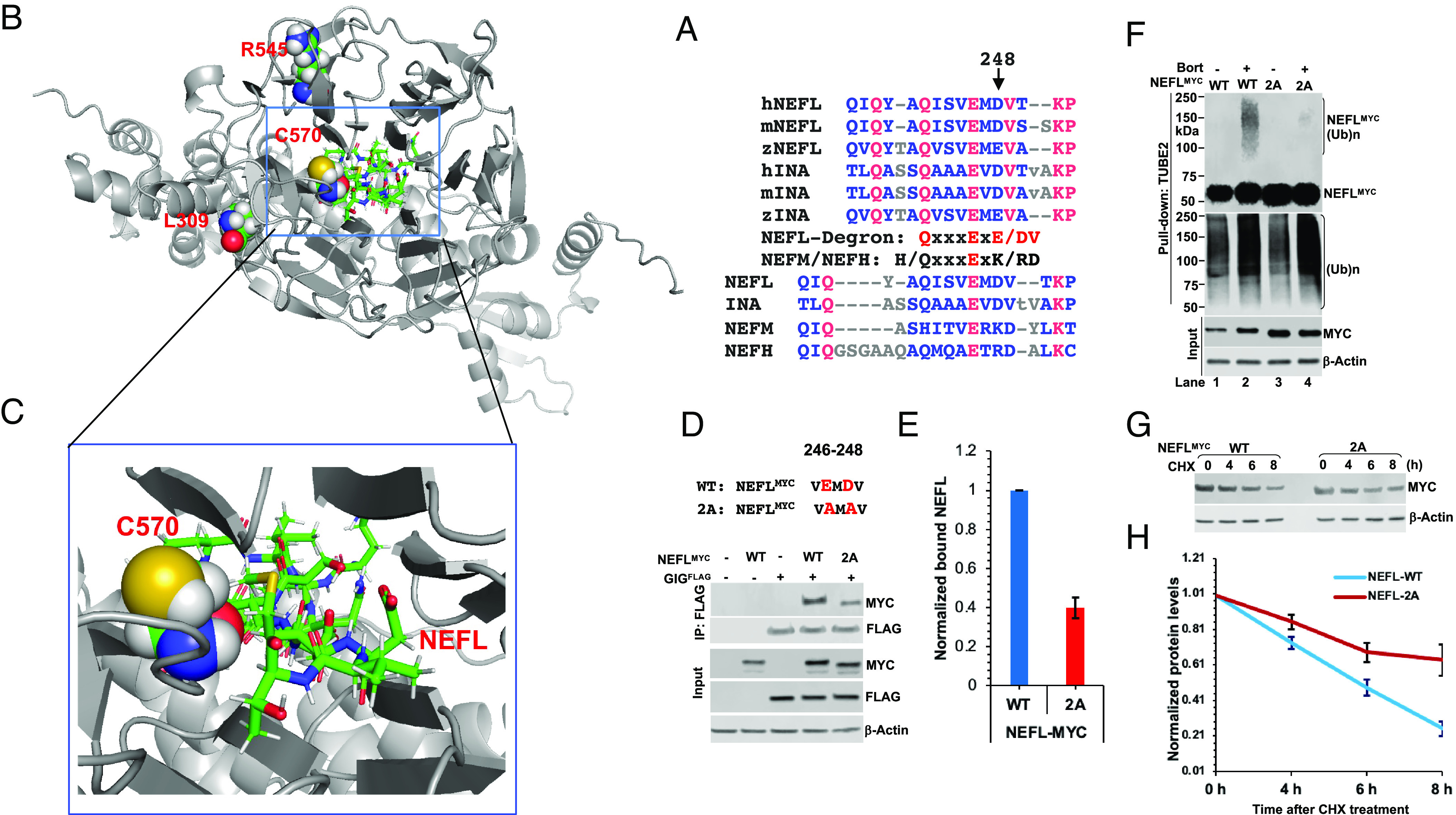
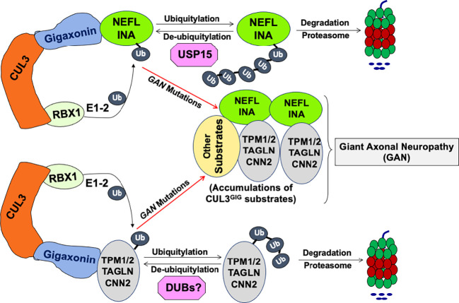
Giant axonal neuropathy (GAN) is caused by mutations in the GAN gene encoding for gigaxonin (GIG), which functions as an adaptor of the CUL3-RBX1-GIG (CRL3GIG) E3 ubiquitin ligase complex. The pathological hallmark of GAN is characterized by the accumulation of densely packed neurofilaments (NFs) in the axons. However, there are fundamental knowledge gaps in our understanding of the molecular mechanisms by which the ubiquitin-proteasome system controls the homeostasis of NF proteins. Recently, the deubiquitylating enzyme USP15 was reported to play a crucial role in regulating ubiquitylation and proteasomal degradation of CRL4CRBN substrate proteins. Here, we report that the CRL3GIG-USP15 pathway governs the destruction of NF proteins NEFL and INA. We identified a specific degron called NEFLL12 degron for CRL3GIG. Notably, mutations in the C-terminal Kelch domain of GIG, represented by L309R, R545C, and C570Y, disrupted the binding of GIG to NEFL and INA, leading to the accumulation of these NF proteins. This accounts for the loss-of-function mutations in GAN patients. In addition to regulating NFs, CRL3GIG also controls actin filaments by directly targeting actin-filament-binding regulatory proteins TPM1, TPM2, TAGLN, and CNN2 for proteasomal degradation. Thus, our findings broadly impact the field by providing fundamental mechanistic insights into regulating extremely long-lived NF proteins NEFL and INA by the CRL3GIG-USP15 pathway and offering previously unexplored therapeutic opportunities to treat GAN patients and other neurodegenerative diseases by explicitly targeting downstream substrates of CRL3GIG.

Keywords: GAN; Gigaxonin; USP15; neurofilament.


Conflict of interest statement

The authors declare no competing interest.

Figures

Fig. 1.
Fig. 1.
USP15 controls the stability of NF proteins. (A) Global proteome analysis of WT and USP15-KO 293FT cells. The threshold of fold changes was set to more than a twofold increase or decrease, and the –Log10 P-value was above 2; two biological and technical replicate samples were subject to mass spectrometric analysis. (B) Validation of proteomic data. WT and USP15-KO 293FT cells were treated with different concentrations of MLN4924 (0, 1, and 2 µM) for 8 h. Cell extracts were analyzed by immunoblotting (IB) with the indicated antibodies. (C) Quantification of NEFL and INA proteins from (B). (D) CHX experiment. USP15+/+ and USP15−/− 293FT cells were treated with cycloheximide (CHX, 50 µg/mL) at the indicated times. Cell extracts were analyzed by IB. (E) Quantification of NEFL and INA proteins from (D). (F) USP15-KO 293FT cells were transiently transfected with empty vector (EV, control), WT USP15Flag, or C298A-USP15Flag mutant plasmid. After 40 h, cells were treated with CHX for the indicated times. Cell extracts were analyzed by IB with the indicated antibodies. (G) Quantification of NEFL, INA, and GS proteins from (F). The relative ratios of indicated proteins:Actin, normalized to time 0, are shown. (H) USP15+/+ and USP15−/− 293FT cells were treated with bortezomib (Bort, 1 µM) for 7 h. Total ubiquitylated proteins were purified using TUBE2-agarose. Bound fractions and input were analyzed by IB. (I) USP15-KO 293FT cells were transiently transfected with NEFLMYC in the presence of EV, WT USP15Flag, or C298A-USP15Flag mutant plasmid. After 40 h, cells were treated with 2 µM bortezomib for 6 h, followed by cell lysis and MYC IP under denaturing conditions. The bound fractions and input were analyzed by IB with antibodies against ubiquitin (Top), MYC, Flag, and Actin. (J) In vitro deubiquitylation assay. USP15−/− 293FT cells were treated with 1 µM Bort for 7 h. Total ubiquitylated proteins, purified using TUBE2, were treated with recombinant (r)USP15 and then analyzed by IB with anti-NEFL and anti-USP15 antibodies. (Ub)n, polyubiquitin. The results shown are representative of three independent experiments (B, D, F, and HJ).
Fig. 2.
Fig. 2.
Depletion of GAN resulted in the accumulation of NF proteins and actin filament–associated regulatory proteins. (A) Comparative proteome analysis of WT (n = 4), GAN-KO2 (n = 3), and GAN-KO16 (n = 3) 293FT cell lines from 10plex TMT analysis. The data are depicted as a volcano plot where both GAN KO clones were combined (n = 6). The red and blue dots denote statistically significant enriched or reduced proteins, respectively. (B) Validation of proteomic data. Cell extracts from WT, GAN-KO2, and GAN-KO16 293FT cell lines were analyzed by IB with the indicated antibodies. (C) Quantification of NEFL, INA, TPM1, TAGLN, CNN2, and BRMS1 proteins from (B). (D) Cell extracts from SH-SY5Y cells, stably expressing shRNA control (shCT) and shRNAs targeting GAN clones 5 and 6 (shGAN_5, shGAN_6), were analyzed by IB with the indicated antibodies. (E) Quantification of NEFL, INA, TPM1, TAGLN, and CNN2 proteins from (D). (F) CHX experiment. WT and GAN-KO2 293FT cells were treated with CHX for the indicated times. Cell extracts were analyzed by IB. (G) Quantification of NEFL, INA, TPM1, TAGLN, CNN2, TPM2, BRMS1, NEFM, and GS proteins from (F). The relative ratios of indicated proteins:Actin, normalized to time 0, are shown. The results shown are representative of at least three independent experiments (B, D, and F).
Fig. 3.
Fig. 3.
CRL3GIG directly ubiquitylates NF proteins and actin filament–associated regulatory proteins. (A) GAN-KO 293FT cells were transfected with the indicated plasmids expressing GIGFLAG and MYC-tagged NEFL (NEFLMYC) for 40 h. Cellular extracts were immunoprecipitated with anti-Flag antibody, followed by IB with antibodies against MYC and FLAG. (B) GAN-KO 293FT cells were transfected with wild-type GIGFLAG. After 48 h, cell extracts were immunoprecipitated with anti-FLAG or IgG control antibody, followed by IB with the indicated antibodies. (C) Endogenous NEFL and INA proteins interact with endogenous GIG. 293FT cells were treated with or without 2 µM MLN4924 for 4 h. Protein extracts were IP with rabbit IgG control or GIG antibody. IP and input samples were analyzed by IB with the indicated antibodies. (D and E) The direct interactions between GST-GIG and NEFL (D) or CNN2 (E) in GST pull-down assays. At least two experimental replicates were repeated to optimize conditions (AE). (F) Cells were treated with or without bortezomib (Bort, 1 µM) for 6 h. Total ubiquitylated proteins were purified using TUBE2-agarose. Bound fractions were analyzed by IB. (G and H) In vitro ubiquitylation reactions of rNEFL (G) and rINA (H) were carried out in the presence or absence of E1, E2, HA-tagged ubiquitin (HAUb) or methylated ubiquitin (Me-Ub), and rCUL3GIG complex. *Indicates a nonspecific band. (I) In vitro competitive ubiquitylation and deubiquitylation assay of NEFL was carried out in the presence of rNEFL, E1, E2, rCUL3GIG complex, ubiquitin mutants with K48 only, K11 only or K63 only and rUSP15. (J) Same as (I), except that reactions were collected at 10 min and 60 min. (KM) In vitro ubiquitylation reactions of rTPM1 (K), rTAGLN (L), and rCNN2 (M). The results shown are representative of at least three independent experiments (FM).
Fig. 4.
Fig. 4.
Mutations in GAN patients at amino acid residues L309, R545, and C570 in the Kelch domain of GIG exhibit a loss-of-function mechanism. (A) Schematic diagram of human GIG. GIG contains BTB, BACK, and Kelch (K1-6) domains. Highly conserved residues in the Kelch domain of GIG across species, which are mutated in GAN patients, including (L309, R545, and C570), are highlighted in red. (B) WT 293FT cells were transfected with empty vector (EV) or wild-type (WT) GIGFLAG and its mutants. After 24 h, cells were treated with 2 µM MLN4924 for 4 h. Cell extracts were immunoprecipitated with anti-FLAG antibody, followed by IB with the indicated antibodies. (C) Quantification of NEFL and INA proteins from (B). The relative ratios of endogenous NEFL and INA interacted with GIGFLAG to input NEFL and INA, normalized to that of WT in lane 2, are shown. (D) Destabilizing endogenous NEFL and INA protein levels in GAN-KO cells by WT GIG, but not its mutants. GAN-KO16 293FT cells were transfected with EV or WT GIGFLAG and its mutants. After 48 h, cell extracts were analyzed by IB with the indicated antibodies. (E) Quantification of NEFL and INA proteins from (D). At least three experimental replicates were repeated to optimize conditions (B and D). (FH) Global proteome analyses (n = 3) of EV-, WT GIGFLAG-, L309R mutant-, and C570Y mutant-transfected GAN-KO 293FT cells. The threshold of fold changes was set to more than 1.41-fold increase or decrease and the –Log10 P-value above 2. The data were depicted as a heatmap (F) and volcano plots for L309R vs. WT (G), and C570Y vs. WT (H). The red and blue dots denote significantly enriched or reduced proteins, respectively.
Fig. 5.
Fig. 5.
Mapping the binding site of GIG on NEFL. (A) Schematic diagram of human NEFL protein. NEFL includes head, rod, and tail domains. The rod domain contains coil 1A, 1B, 2A, and 2B subdomains, separated by linkers L1, L12, and L2. Deletion of amino acids 237–279 (D4) highlighted in red abolished the binding to GIG, indicating that this sequence contains the NEFL degron recognized by GIG. The sequence alignment of L12, 2A, and L2 segments from human NEFL, INA, NEFM, and NEFH was shown. (B) 293FT cells were transfected with the indicated plasmids expressing GIGFLAG and full-length (FL) or deletions of NEFLMYC for 40 h. Cellular extracts were immunoprecipitated with anti-Flag antibody, followed by IB with antibodies against MYC and FLAG. A band ~ 25 kDa (top panel) represents IgG light chains (IgG-LC). (C) Design of NEFL-L12, -2A, -L2 and NEFM-L12 (M-L12) peptides. (D) Peptide pull-down assays were performed using GSTGIG protein and NEFL-L12, -2A, and –L2 peptides (lane 2 without peptide used as negative control), followed by analyzed by IB with anti-GST antibody. (E) Same as (D), except that GIGFLAG protein and NEFM-L12 (M-L12) were used. Data are representative of three independent experiments (B, D, and E).
Fig. 6.
Fig. 6.
Identification of molecular basis for the specific recognition of NEFL by GIG. (A) L12 segments from human (h), mouse (m), and zebrafish (z) NEFL and INA orthologs and from human NEFL, INA, NEFM, and NEFH. (B) Docking pose of the NEFLL12 degron to GIG (n = 10). The NEFLL12 degron binds to the Kelch domain of GIG and directly interacts with residue C570 located close to residues L309 and R545. (C) A close view of electrostatic interactions between residue C570 and the motif ExD. (D) WT 293FT cells, transfected with the indicated plasmids expressing GIGFLAG and NEFLMYC WT or 2A mutant for 24 h, were treated with 2 µM MLN4924 for 4 h. Cell extracts were immunoprecipitated with anti-Flag antibody, followed by IB with antibodies against MYC and FLAG. (E) Quantification of NEFLMYC protein interacted with GIGFLAG from (D). Error bars represent ±SD; n = 2. (F) 293FT cells, transfected with NEFLMYC WT or 2A mutant for 40 h, were treated with bortezomib (Bort, 2 µM) for 7 h. Total ubiquitylated proteins were purified using TUBE2-agarose, followed by IB with anti-MYC and anti-ubiquitin antibodies. (Ub)n, polyubiquitin. (G) Same as (F), except that cells were treated with CHX at the indicated times. Cell extracts were analyzed by IB with anti-MYC and Actin. (H) Quantification of NEFL-MYC protein from (G). Error bars represent ±SD; n = 2.
Fig. 7.
Fig. 7.
Proposed model: the CRL3GIG–USP15 pathway governs the destruction of neurofilament proteins NEFL and INA. CRL3GIG also regulates the ubiquitylation of actin filament–associated regulatory proteins (TPM1, TPM2, TAGLN, and CNN2), which DUBs may deubiquitylate. Mutations in the GAN gene encoding Gigaxonin cause giant axonal neuropathy (GAN), characterized by abnormal accumulation of intermediate filaments in both neuronal and non-neuronal cells. Thus, CRL3GIG may regulate a vast repertoire of substrates. For the sake of simplicity, the homodimerization of Gigaxonin is omitted.

References

    1. Varshavsky A., The ubiquitin system, an immense realm. Annu. Rev. Biochem. 81, 167–176 (2012). - PubMed
    1. Hershko A., Ciechanover A., The ubiquitin system. Annu. Rev. Biochem. 67, 425–479 (1998). - PubMed
    1. Huang X., Dixit V. M., Drugging the undruggables: Exploring the ubiquitin system for drug development. Cell Res. 26, 484–498 (2016). - PMC - PubMed
    1. Petroski M. D., Deshaies R. J., Function and regulation of cullin-RING ubiquitin ligases. Nat. Rev. Mol. Cell Biol. 6, 9–20 (2005). - PubMed
    1. Harper J. W., Schulman B. A., Cullin-RING ubiquitin ligase regulatory circuits: A quarter century beyond the F-Box hypothesis. Annu. Rev. Biochem. 90, 403–429 (2021). - PMC - PubMed

Publication types