

Diagnosis of an aortico-left ventricular tunnel in a fetus: a case report
- PMID: 37903312
- PMCID: PMC10617269
- DOI: 10.1177/03000605231207756
Diagnosis of an aortico-left ventricular tunnel in a fetus: a case report
Abstract
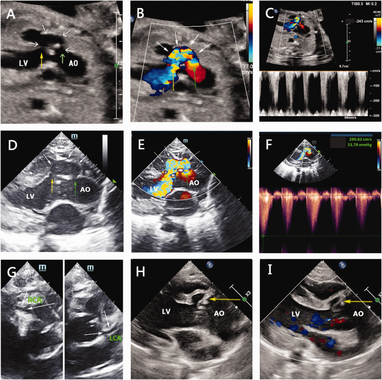
An aortico-left ventricular tunnel is a rare congenital heart disease, and its prenatal diagnosis is even rarer. This report describes a fetus diagnosed with an aortico-left ventricular tunnel at 26 weeks of gestation. After delivery, the infant exhibited cyanosis and cessation of breathing. After resuscitation, he was transferred to the neonatal intensive care unit. Echocardiography confirmed an aortico-left ventricular tunnel. The infant survived after surgical repair. An aortico-left ventricular tunnel can be diagnosed by antenatal ultrasound, and prompt neonatal management can help to prevent perinatal morbidity and mortality.
Keywords: Aortico-left ventricular tunnel; case report; echocardiography; fetus; neonatal; prenatal diagnosis.
Conflict of interest statement
The authors have no conflicts of interest to disclose.
Figures
Similar articles
-
Aortico-right ventricular tunnel: prenatal diagnosis leading to neonatal survival.Fetal Diagn Ther. 2007;22(5):335-8. doi: 10.1159/000103291. Epub 2007 Jun 5. Fetal Diagn Ther. 2007. PMID: 17556819
-
Aortico-left ventricular tunnel in fetuses and infants.Ann Thorac Surg. 1996 Jun;61(6):1805-10. doi: 10.1016/0003-4975(96)00189-0. Ann Thorac Surg. 1996. PMID: 8651788
-
Prenatal diagnosis of an aortico-left ventricular tunnel.Ultrasound Obstet Gynecol. 2000 May;15(5):435-8. doi: 10.1046/j.1469-0705.2000.00119.x. Ultrasound Obstet Gynecol. 2000. PMID: 10976489
-
[Aortico-left ventricular tunnel. Report of 3 cases and review of the literature].Z Kardiol. 1982 Oct;71(10):695-704. Z Kardiol. 1982. PMID: 6760571 Review. German.
-
Repair of aortico-right ventricular tunnel.Eur J Cardiothorac Surg. 1998 Aug;14(2):214-7. doi: 10.1016/s1010-7940(98)00168-7. Eur J Cardiothorac Surg. 1998. PMID: 9755011 Review.
References
-
- Edwards JE. An atlas of acquired disease of the heart and great vessels. 2nd. Vol. 2. WB Saunders; Philadelphia: 1961. p. 1142.
-
- Levy MJ, Schachner A, Blieden LC. Aortico-left ventricular tunnel: collective review. J Thorac Cardiovasc Surg 1982; 84: 102–109. 1982/07/01. - PubMed
-
- Chen LL, Zhang CQ, Gong LG. A special type aortico-left ventricular tunnel with bicuspid aortic valve. Int J Cardiovasc Imaging 2020; 36: 55–57. 2019/09/11. DOI: 10.1007/s10554-019-01692-9. - PubMed
Publication types
MeSH terms
LinkOut - more resources
Full Text Sources
Medical
