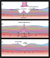
RETINAL OPTICAL COHERENCE TOMOGRAPHY IMAGING BIOMARKERS: A Review of the Literature
- PMID: 37903455
- PMCID: PMC10885864
- DOI: 10.1097/IAE.0000000000003974
RETINAL OPTICAL COHERENCE TOMOGRAPHY IMAGING BIOMARKERS: A Review of the Literature
Abstract
Purpose: The aim of this literature review was to summarize novel optical coherence tomography (OCT) imaging biomarkers that have recently been described in the literature and are frequently encountered clinically.
Methods: The literature was reviewed to identify novel OCT biomarkers reported to date. A descriptive summary of all terms and representative illustrations were provided to highlight the most relevant features.
Results: Thirty-seven OCT terminologies were identified. The vitreomacular interface disorder group included the four stages of epiretinal membrane, macular pseudohole, tractional lamellar hole (LH), degenerative LH, cotton ball sign, and foveal crack sign. The age-related macular degeneration group included outer retinal tubulation, multilayered pigment epithelial detachment, prechoroidal cleft, onion sign, double-layer sign, complete outer retinal atrophy, complete retinal pigment epithelium and outer retinal atrophy, and reticular pseudodrusen. The uveitic disorder group consisted of bacillary layer detachment, syphilis placoid, rain-cloud sign, and pitchfork sign. The disorders relating to the toxicity group included flying saucer sign and mitogen-activated protein kinase (MEK) inhibitor-associated retinopathy. The disorders associated with the systemic condition group included choroidal nodules and needle sign. The pachychoroid spectrum group included pachychoroid and brush border pattern. The vascular disorder group included pearl necklace sign, diffuse retinal thickening, disorganization of retinal inner layers, inner nuclear layer microcysts, hyperreflective retinal spots, paracentral acute middle maculopathy, and acute macular neuroretinopathy. The miscellaneous group included omega sign (ω), macular telangiectasia (type 2), and omega sign (Ω).
Conclusions: Thirty-seven OCT terminologies were summarized, and detailed illustrations consolidating the features of each biomarker were included. A nuanced understanding of OCT biomarkers and their clinical significance is essential because of their predictive and prognostic value.
Copyright © 2023 The Author(s). Published by Wolters Kluwer Health, Inc. on behalf of the Opthalmic Communications Society, Inc.
Conflict of interest statement
None of the authors has any conflicting interests to disclose.
Figures


References
-
- Sun JK, Lin MM, Lammer J, et al. . Disorganization of the retinal inner layers as a predictor of visual acuity in eyes with center-involved diabetic macular edema. JAMA Ophthalmol 2014;132:1309–1316. - PubMed
-
- Ishibashi T, Iwama Y, Nakashima H, et al. . Foveal Crack Sign: an OCT sign preceding macular hole after vitrectomy for rhegmatogenous retinal detachment. Am J Ophthalmol 2020;218:192–198. - PubMed
-
- Govetto A, Lalane RA, 3rd, Sarraf D, et al. . Insights into epiretinal membranes: presence of ectopic inner foveal layers and a new optical coherence tomography staging scheme. Am J Ophthalmol 2017;175:99–113. - PubMed
-
- Govetto A, Virgili G, Rodriguez FJ, et al. . Functional and anatomical significance of the ectopic inner foveal layers in eyes with idiopathic epiretinal membranes: surgical results at 12 months. Retina 2019;39:347–357. - PubMed
Publication types
MeSH terms
Substances
LinkOut - more resources
Full Text Sources

