. 2023 Oct 30;14(1):6908.
doi: 10.1038/s41467-023-42702-8.

Farnesoid X receptor activation by bile acids suppresses lipid peroxidation and ferroptosis

Affiliations

Farnesoid X receptor activation by bile acids suppresses lipid peroxidation and ferroptosis

Juliane Tschuck et al. Nat Commun. .

Abstract

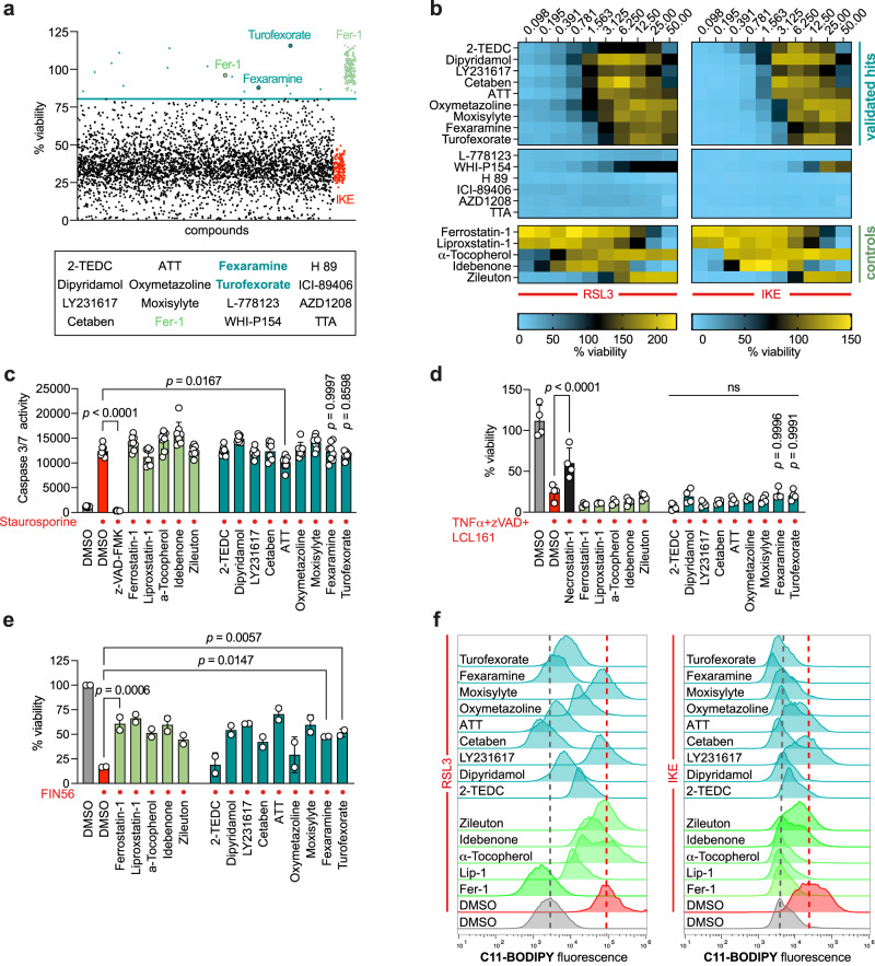
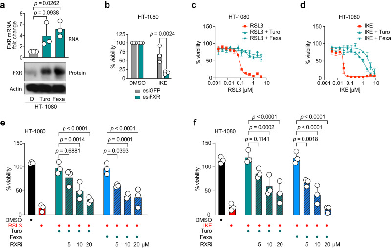
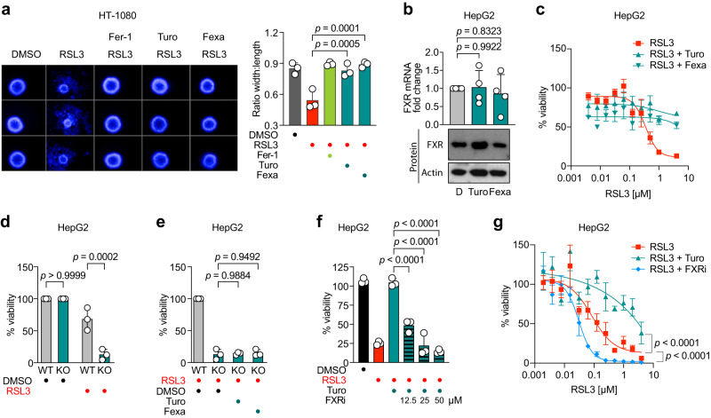
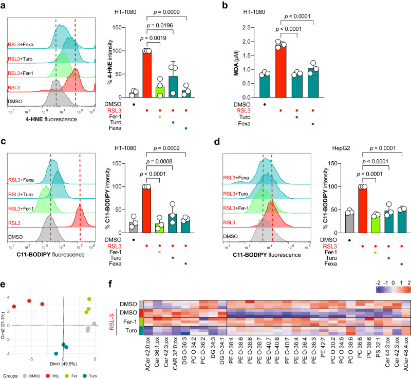
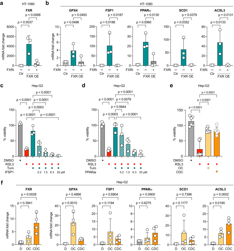
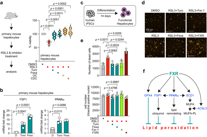
Ferroptosis is a regulated cell death modality that occurs upon iron-dependent lipid peroxidation. Recent research has identified many regulators that induce or inhibit ferroptosis; yet, many regulatory processes and networks remain to be elucidated. In this study, we performed a chemical genetics screen using small molecules with known mode of action and identified two agonists of the nuclear receptor Farnesoid X Receptor (FXR) that suppress ferroptosis, but not apoptosis or necroptosis. We demonstrate that in liver cells with high FXR levels, knockout or inhibition of FXR sensitized cells to ferroptotic cell death, whereas activation of FXR by bile acids inhibited ferroptosis. Furthermore, FXR inhibited ferroptosis in ex vivo mouse hepatocytes and human hepatocytes differentiated from induced pluripotent stem cells. Activation of FXR significantly reduced lipid peroxidation by upregulating the ferroptosis gatekeepers GPX4, FSP1, PPARα, SCD1, and ACSL3. Together, we report that FXR coordinates the expression of ferroptosis-inhibitory regulators to reduce lipid peroxidation, thereby acting as a guardian of ferroptosis.


Conflict of interest statement

J.T. and K.H. are inventors on a patent application involving ferroptosis. The remaining authors declare no competing interests.

Figures

Fig. 1
Fig. 1. Identification and validation of novel ferroptosis inhibitors.
a Viability screen of 3684 small molecules (see Supplementary Data 1) identified Turofexorate and Fexaramine among 13 other hits as ferroptosis inhibitors after induction with 1.5 µM IKE for 18 h. 2 µM Ferrostatin-1 (Fer-1) was used as positive control. Hit threshold was set as 3x SD from median of compound-treated wells. b Dose-response heat map of cell viability with primary hits after ferroptosis induction with 100 nM RSL3 or 1.5 µM IKE for 18 h (n = 3 technical replicates). Known ferroptosis inhibitors served as positive controls. c Apoptotic cell death was induced with 1 µM Staurosporine in HT-1080 for 18 h, 50 µM Z-VAD-FMK was used as a known apoptosis inhibitor. Compound concentrations can be found in Supplementary Data 2; one-way ANOVA with Dunnett’s test. Data are mean ± SD of n = 8 technical replicates. d Necroptotic cell death was induced with 20 ng/ml TNFα + 10 µM Z-VAD-FMK + 10 µM LCL161 for 18 h in MEFs, 10 µM Necrostatin-1 was used to inhibit necroptosis. Compound concentrations can be found in Supplementary Data 2; one-way ANOVA with Dunnett’s test. Data are mean ± SD of n = 4 technical replicates. e Turofexorate and Fexaramine inhibit FIN56-induced cell death in HT-1080. Ferroptosis was induced by 200 nM FIN56 for 18 h. Compound concentrations can be found in Supplementary Data 2; one-way ANOVA with Dunnett’s test. Data are mean ± SD of n = 2 technical replicates. f Selected compounds inhibit lipid peroxidation in C11-BODIPY-stained cells. Ferroptosis was induced by 250 nM RSL3 for 2 h, or 2.5 µM IKE for 6 h, compound concentrations can be found in Supplementary Data 2.
Fig. 2
Fig. 2. Activation of FXR suppresses ferroptosis in cooperation with RXR.
a Treatment of HT-1080 with 12 µM Turofexorate or Fexaramine for 7 h increases FXR expression. Levels of mRNA were normalized to GAPDH expression. Data are mean ± SD of n = 3 biological replicates. Western Blot shown is one representative experiment from n = 3. One-way ANOVA with Dunnett’s test; D = DMSO b Knockdown of FXR sensitizes cells to ferroptosis. HT-1080 were transfected with 40 nM esiRNA against FXR for 48 h and treated with 1 µM IKE for 18 h. EsiGFP-transfected cells served as a control group. Data are mean ± SD of n = 3 biological replicates; one-way ANOVA with Tukey’s test. c, d Turofexorate and Fexaramine are able to rescue RSL3- and IKE-induced ferroptosis. Cells were treated with 12 µM Turofexorate or Fexaramine and indicated dose range of inducers (RSL3 and IKE) for 18 h. Data are normalized to DMSO and plotted as mean ± SD of n = 3 biological replicates. e, f Dimerization of FXR with RXR is necessary to conduct anti-ferroptotic effect. Ferroptosis was induced with 250 nM RSL3, or 1 µM IKE for 18 h, and 12 µM Turofexorate or Fexaramine was used to inhibit ferroptosis. The RXR transactivation inhibitor HX 531 (RXRi) was added in indicated doses. Data are normalized to untreated control and plotted as mean ± SD of n = 3 biological replicates with each 6 technical replicates; one-way ANOVA with Tukey’s test.
Fig. 3
Fig. 3. FXR suppresses ferroptosis in 3D spheroid and hepatic cell models.
a HT-1080 3D spheroid models are rescued from ferroptosis by treatment with 12 µM Turofexorate or Fexaramine for 48 h. Ferroptosis was induced by 200 nM RSL3 for 48 h, 2 µM Fer-1 was used as a positive control. Representative spheroid images are shown. Mean data ± SD from n = 3 biological replicates with each 8 spheroids were quantified by high-content image analysis; one-way ANOVA with Dunnett’s test. b HepG2 cells treated with 12 µM Turofexorate or Fexaramine do not show increased levels of FXR. Levels of mRNA were normalized to GAPDH expression. Data plotted are mean ± SD of n = 4 biological replicates. Western Blot shown is one representative experiment from n = 3. One-way ANOVA with Dunnett’s test; D = DMSO. c HepG2 cells have lower ferroptosis sensitivity when treated with 250 nM RSL3, but ferroptosis can be rescued by 12 µM Turofexorate or Fexaramine. Ferroptosis was induced for 18 h. Data plotted are mean ± SD of n = 3 biological replicates. d HepG2 cells with FXR knockout show a higher sensitivity towards ferroptosis induction. Cells were treated with 125 nM RSL3 for 18 h. Data are normalized to DMSO-treated control and plotted as mean ± SD of n = 3 biological replicates; 2-way-ANOVA with Šídák’s test. e HepG2 cells with FXR KO could not be rescued from ferroptosis by treatment with 12 µM Turofexorate or Fexaramine. Ferroptosis was induced with 125 nM RSL3 for 18 h. Data are normalized to wildtype cells treated with 125 nM RSL3 and plotted as mean ± SD of n = 3 biological replicates; one-way ANOVA with Tukey’s test. f Ferroptosis was induced with 250 nM RSL3 for 18 h, and 12 µM Turofexorate was used to inhibit ferroptosis. The FXR inhibitor Guggulsterone (FXRi) was added in indicated doses. Data are normalized to untreated control and plotted as mean ± SD of n = 3 biological replicates; one-way ANOVA with Tukey’s test. g HepG2 cells can be sensitized towards ferroptosis (250 nM RSL3) by FXR inhibitor Guggulsterone (FXRi). Ferroptosis was induced for 18 h, 12 µM Turofexorate and 50 µM Guggulsterone were used. Data are mean ± SEM of n = 3 biological replicates; 2-way-ANOVA with Šídák’s test.
Fig. 4
Fig. 4. Activation of FXR reduces lipid peroxidation.
a FXR activation by 12 µM Turofexorate or Fexaramine reduces 4-HNE levels. Ferroptosis was induced by 300 nM RSL3 for 2 h. 2 µM Fer-1 was used as positive control. Representative flow cytometry histogram is shown, Data plotted are % intensity of median fluorescence normalized to RSL3 treated cells ± SD of n = 3 biological replicates; one-way ANOVA with Tukey’s test. b FXR activation reduces MDA levels in TBARS assay. Ferroptosis was induced with 250 nM RSL3 for 2.5 h, cells were rescued with 12 µM Turofexorate or Fexaramine. Data are mean ± SD of n = 3 biological replicates; one-way ANOVA with Tukey’s test. c, d FXR activation inhibits lipid peroxidation in HT-1080 and HepG2 stained with BODIPY-C11 lipid peroxidation sensor. Cells were treated with 250 nM RSL3 and 12 µM Turofexorate or Fexaramine for 2 h. 2 µM Fer-1 served as a positive control. Representative flow cytometry histograms are shown. Data plotted are % intensity of median fluorescence normalized to RSL3 treated cells ± SD of n = 3 biological replicates; one-way ANOVA with Tukey’s test. e PCA illustrating the phenotypic differences in the cellular pool of HT-1080 cells treated with RSL3, DMSO, RSL3+Turofexorate, and RSL3+Ferrostatin-1 (n = 3 technical replicates). f HT-1080 cells treated with 200 nM RSL3 for 2 h show modified lipids. PUFA (polyunsaturated fatty acid) containing phospholipids are the predominate drivers. We observed a decrease in relevant highly unsaturated phospholipid species during ferroptosis (RSL3) initiation. Ferrostatin-1 (2 µM) and Turofexorate (12 µM) protected these lipids (n = 3 technical replicates). ACer Acylceramide, Cer Ceramide, CAR Carnitine, DG Diacylglycerol, PC Phosphatidylcholine, PE Phosphatidylethanolamine, PS Phosphatidylserine.
Fig. 5
Fig. 5. FXR activation induces expression of ferroptosis-inhibitory genes.
a Overexpression of FXR in HT-1080. Levels of mRNA were normalized to GAPDH mRNA. FXR levels are reverted to control levels upon treatment with 50 µM FXR inhibitor Guggulsterone for 24 h. Data are mean ± SD of n = 3 biological replicates; one-way ANOVA with Dunnett’s test. b FXR overexpression leads to elevated mRNA levels of anti-ferroptotic genes GPX4, FSP1, PPARα, ACSL3 and SCD1. Expression levels are normalized to mRNA levels of GAPDH. Upregulated anti-ferroptotic target genes in HT-1080 overexpressing FXR can be reverted to control levels by treatment with 50 µM FXR inhibitor Guggulsterone for 24 h. RP2 was used as a housekeeper gene for normalization. Data are mean ± SD of n = 3 biological replicates; one-way ANOVA with Dunnett’s test. c, d Ferroptosis inhibition by FXR activation is reverted when FSP1 or PPARα are inhibited. Ferroptosis was induced by 200 nM RSL3 for 18 h, 12 µM Turofexorate was used to inhibit ferroptosis. FSP1 was inhibited by iFSP1, PPARα was inhibited by GW6471 (PPARαi). Data are normalized to untreated control and plotted as mean ± SD of n = 3 biological replicates; one-way ANOVA with Tukey’s test. e Endogenous FXR activation by bile acids inhibits ferroptosis in HepG2 cells. Cells were treated with 65 nM RSL3, 20 µM Chenodeoxycholic acid (CDC) and 2 µM Obeticholic acid (OC) for 5 h. Data are mean ± SD of n = 6 biological replicates; one-way ANOVA with Dunnett’s test. f Levels of FXR and anti-ferroptotic genes GPX4, FSP1, PPARα, ACSL3 and SCD1 are elevated after bile acid treatment of HepG2. Cells were treated with 20 µM Chenodeoxycholic acid (CDC) or 2 µM Obeticholic acid (OC) for 8 h. Expression levels are normalized to mRNA levels of GAPDH. Data are mean ± SD of n = 4 biological replicates; one-way ANOVA with Dunnett’s test; D = DMSO.
Fig. 6
Fig. 6. FXR activation inhibits ferroptosis in primary mouse hepatocytes and human iPSC-derived hepatocytes.
a Activation of FXR inhibits RSL3-induced ferroptosis in primary mouse hepatocytes. Isolated cells were treated with 1 µM RSL3 and 12 µM Turofexorate/Fexaramine, 2 µM OC or 20 µM CDC for 18 h; n = 3 biological replicates; one-way ANOVA with Dunnett’s test. b Levels of FSP1 and PPARα are elevated after 12 µM Turofexorate and Fexaramine treatment of primary mouse hepatocytes. Expression levels are normalized to mRNA levels of GAPDH. Data are mean ± SD of n = 3 biological replicates; one-way-ANOVA with Dunnett’s test. D = DMSO. c Induced pluripotent stem cells were differentiated into functional hepatocytes and treated with RSL3 to generate a human primary hepatocyte cell model. d Ferroptosis was induced in differentiated hepatocytes (less sensitive) by treatment with 1 µM RSL3 for 6 h. To rescue cells from ferroptosis, cells were co-treated with 12 µM Turofexorate or Fexaramine or 2 µM Ferrostatin-1. To sensitize cells to RSL3-induced ferroptosis, cells were treated with 50 µM FXR inhibitor (FXRi). Hepatocytes were stained with 5 µg/ml Propidium iodide for live/dead discrimination; scale bar is 200 µm. e Quantification of PI-stained hepatocytes. Cotreatment with Turofexorate or Fexaramine rescues cells from ferroptosis, whereas inhibition of FXR sensitizes to cell death. Normalization of cell numbers was done by Hoechst staining. Data are mean ± SD of n = 3–4 biological replicates; one-way ANOVA with Dunnett’s test. f Graphical summary of ferroptosis inhibition by FXR activation.

References

    1. Galluzzi L, et al. Molecular mechanisms of cell death: recommendations of the Nomenclature Committee on Cell Death 2018. Cell Death Differ. 2018;25:486–541. doi: 10.1038/s41418-017-0012-4. - DOI - PMC - PubMed
    1. Hadian K, Stockwell BR. The therapeutic potential of targeting regulated non-apoptotic cell death. Nat. Rev. Drug Discov. 2023;22:723–742. doi: 10.1038/s41573-023-00749-8. - DOI - PubMed
    1. Dixon SJ, et al. Ferroptosis: an iron-dependent form of nonapoptotic cell death. Cell. 2012;149:1060–1072. doi: 10.1016/j.cell.2012.03.042. - DOI - PMC - PubMed
    1. Hadian K, Stockwell BR. SnapShot: Ferroptosis. Cell. 2020;181:1188–1188. doi: 10.1016/j.cell.2020.04.039. - DOI - PMC - PubMed
    1. Jiang X, Stockwell BR, Conrad M. Ferroptosis: mechanisms, biology and role in disease. Nat. Rev. Mol. Cell Biol. 2021;22:266–282. doi: 10.1038/s41580-020-00324-8. - DOI - PMC - PubMed

Publication types

Substances