. 2023 Oct 31;9(1):402.
doi: 10.1038/s41420-023-01697-3.

Adropin inhibits the progression of atherosclerosis in ApoE-/-/Enho-/- mice by regulating endothelial-to-mesenchymal transition

Affiliations

Adropin inhibits the progression of atherosclerosis in ApoE-/-/Enho-/- mice by regulating endothelial-to-mesenchymal transition

Teng Ying et al. Cell Death Discov. .

Abstract

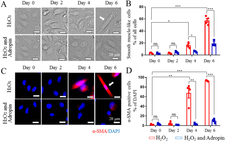
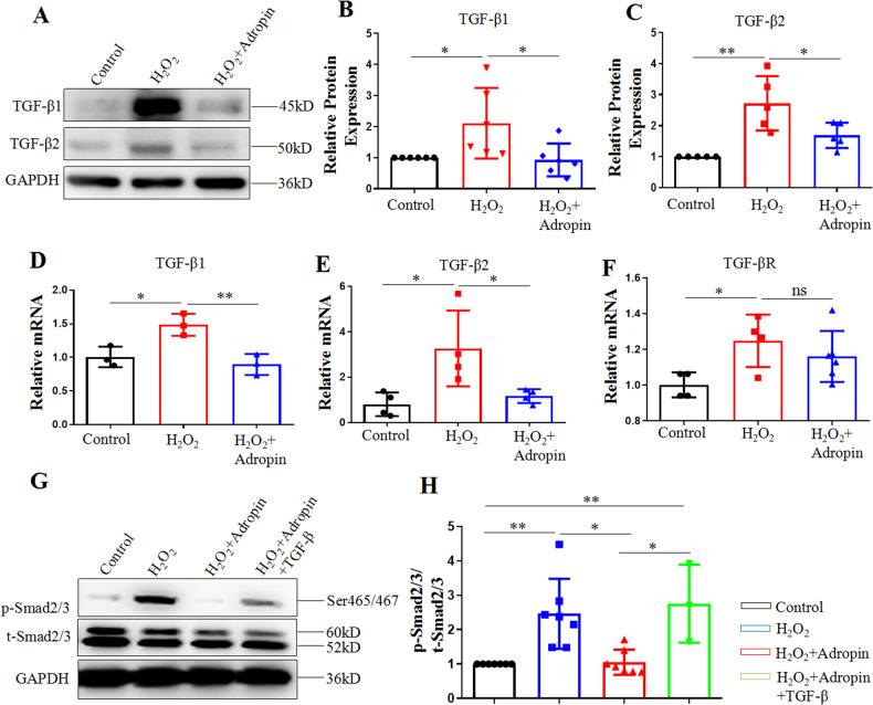
Adropin, a secreted protein, coded by energy homeostasis-associated gene (Enho), is recently reported to modulate atherogenesis, with endothelial-to-mesenchymal transition (EndMT) involved in the early process. We explored whether adropin may alleviate atherosclerosis by regulating EndMT. We found that an intraperitoneal injection of adropin [105 μg/(kg·d) for 13 weeks] inhibited the progression of high-fat diet (HFD)-induced aortic atherosclerosis in apolipoprotein E-deficient mice (ApoE-/-) and those with double gene deletion (ApoE-/-/Enho-/-), as detected by Oil Red O and haematoxylin-eosin staining. In the aortas of ApoE-/- mouse, adropin treatment ameliorated the decrease in the mRNA expression of endothelial cell markers (leukocyte differentiation antigen 31, CD31, and vascular endothelial cadherin, VE-cadherin), but increased that of EndMT markers (alpha smooth muscle actin, α-SMA, and fibroblasts specific protein-1). In vitro, an adropin treatment (30 ng/ml) arrested the hydrogen peroxide (H2O2)-induced EndMT in human umbilical vein endothelial cells (HUVECs), attenuated the morphological changes of HUVECs, reduced the number of immunofluorescence-positive α-SMA, increased the mRNA and protein expressions of CD31 and VE-cadherin, and decreased those of α-SMA. Furthermore, the adropin treatment decreased the mRNA and protein expressions of transforming growth factor (TGF)-β1 and TGF-β2, and suppressed the phosphorylation of downstream signal protein Smad2/3 in HUVECs. These mitigative effects of adropin on H2O2-induced EndMT were reversed by the transfection of TGF-β plasmid. The findings signify that adropin treatment may alleviate the atherosclerosis in ApoE-/-/Enho-/- mice by inhibiting EndMT via the TGF-β/Smad2/3 signaling pathway.


Conflict of interest statement

The authors declare no competing interests.

Figures

Fig. 1
Fig. 1. The alleviated atherosclerosis in ApoE-/- mice and ApoE-/-/Enho-/- mice by adropin treatment.
A Cross-sections of the aortic roots were stained by ORO, red part showing lipid in plaque. B H&E staining from the same samples by ORO staining, revealing plaque tissues protruding from lumen. C Gross morphology of the whole aortas, the red parts indicating lipid plaque by ORO staining. D The result of ORO staining indicated as a percentage of lipid area/plaque area. E The result of H&E staining was shown as a percentage of plaque area/lumen area. F The result of ORO staining in the whole aortas was shown as a percentage of lipid plaque area/aorta area. The above results revealed that the aggravated atherosclerosis in ApoE-/- mice or ApoE-/-/Enho-/- mice fed with high fat diet was reversed by the adropin treatment. ND, the normal diet group. HFD, the high fat diet group. HFD+Adropin, the group fed with HFD together with Adropin treatment. DKO-HFD group: ApoE-/-/Enho-/- double-gene-knockout (DKO) mice were fed with HFD. DKO-HFD+Adropin group: DKO mice were fed with HFD together with Adropin. H&E, haematoxylin-eosin. The data are presented as mean ± SEM. and analyzed by ANOVA. n = 5. *P < 0.05, as compared with ND; # P < 0.05, as compared with HFD; & P < 0.05, as compared with DKO-HFD. Bar = 500 μm in (A) and (B), and 10 mm in C.
Fig. 2
Fig. 2. The qualitative and quantitative analyses of endothelial-to-mesenchymal transition (EndMT).
A The leftmost column: a complete view of cross-sections of the aortic roots, with the rectangle indicating the sites where the amplified pictures in other four column came from. The left second column (DAPI, blue): the nuclei of 4’,6-diamidino-2-phenylindole positive cells. The median column (endothelial cell marker CD31, red): the complete expression of CD31 was showed in aortic intima (arrowhead) in the ND group, and HFD led to absence in the expression of CD31, which was rescued by adropin treatment (the HFD+Adropin group). The right second column (mesenchymal cell marker SM22α, green): oval box showed that HFD induced obvious plaque formation and mass expression of SM22α with no obvious CD31 expression in aortic intima, indicating the occurrence of EndMT, which was reversed by adropin treatment. BE The statistical histograms of mRNA expressions from endothelial cell markers CD31 and VE-cadherin, and mesenchymal cell markers α-SMA and FSP-1. The rightmost column: merged images. CD31, leukocyte differentiation antigen 31. VE-cadherin, vascular endothelial cadherin. α-SMA, alpha smooth muscle actin. FSP, fibroblasts specific protein-1. SM22α, smooth muscle 22 alpha. DAPI, 4’,6-diamidino-2-phenylindole. Bar = 500 μm. The data are presented as mean ± SEM. and analyzed by ANOVA. n = 3–5. *P < 0.05, **P < 0.01, ***P < 0.001.
Fig. 3
Fig. 3. The inhibited hydrogen peroxide (H2O2)-induced conversion of HUVECs into smooth muscle cells (SMCs) by adropin treatment.
A H2O2-induced morphological changes of HUVECs at different timepoints. Fusiform cells were considered as smooth muscle-like cells after 4 days of culture (arrowhead). B The percentage of fusiform smooth muscle-like cells in the total HUVECs. C H2O2-induced α-SMA positive expression in HUVECs at different timepoints by immunofluorescence. Fusiform α-SMA positive cells were marked red (arrowhead). D The percentage of α-SMA positive cells (red) /DAPI positive cells (blue). Adropin treatment (the H2O2 + Adropin) alleviated H2O2-induced SMCs-like change (A, B) and expression of mesenchymal cell marker α-SMA (C, D) in HUVECs, suggesting the reversal of EndMT. HUVECs, human umbilical vein endothelial cells. α-SMA, alpha smooth muscle actin. DAPI, 4’,6-diamidino-2-phenylindole. EndMT, Endothelial-to-mesenchymal transition. The data are presented as mean ± SEM. and analyzed by t tests. n = 4. *P < 0.05, **P < 0.01, ***P < 0.001. Bar = 20 μm.
Fig. 4
Fig. 4. The maintained gene and protein expressions of CD31,VE-cadherin TGF-β1 and α-SMA in HUVECs by adropin treatment.
AC Western blotting results of CD31 and VE-cadherin. D, E q-PCR results of CD31 and VE-cadherin. F, G Western blotting results of α-SMA. H q-PCR results of TGF-β1. I q-PCR results of α-SMA. The data are presented as mean ± SEM. and analyzed by ANOVA. n = 3–7. *P < 0.05, **P < 0.01, *P < 0.001.
Fig. 5
Fig. 5. The inhibited TGF-β/Smad2/3 pathway in vitro in HUVECs by adropin treatment.
AC Western blotting results of TGF-β1 and TGF-β2. D, E q-PCR results of TGF-β1 and TGF-β2. F q-PCR results of TGF-βR. G, H Western blotting results of total and phosphorylated Smad2/3. HUVECs, human umbilical vein endothelial cells. TGF-β, transforming growth factor-β. TGF-βR, transforming growth factor-β receptor. The data are presented as mean ± SEM. and analyzed by ANOVA. *P < 0.05, **P < 0.01.

References

    1. Moonen JR, Lee ES, Schmidt M, Maleszewska M, Koerts JA, Brouwer LA, et al. Endothelial-to-mesenchymal transition contributes to fibro-proliferative vascular disease and is modulated by fluid shear stress. Cardiovasc Res. 2015;108:377–86. doi: 10.1093/cvr/cvv175. - DOI - PubMed
    1. Chen PY, Qin L, Baeyens N, Li G, Afolabi T, Budatha M, et al. Endothelial-to-mesenchymal transition drives atherosclerosis progression. J Clin Investig. 2015;125:4514–28. doi: 10.1172/JCI82719. - DOI - PMC - PubMed
    1. Evrard SM, Lecce L, Michelis KC, Nomura-Kitabayashi A, Pandey G, Purushothaman KR, et al. Endothelial to mesenchymal transition is common in atherosclerotic lesions and is associated with plaque instability. Nat Commun. 2016;7:11853. doi: 10.1038/ncomms11853. - DOI - PMC - PubMed
    1. Krenning G, Moonen JR, van Luyn MJ, Harmsen MC. Vascular smooth muscle cells for use in vascular tissue engineering obtained by endothelial-to-mesenchymal transdifferentiation (EnMT) on collagen matrices. Biomaterials. 2008;29:3703–11. doi: 10.1016/j.biomaterials.2008.05.034. - DOI - PubMed
    1. Moonen JR, Krenning G, Brinker MG, Koerts JA, van Luyn MJ, Harmsen MC. Endothelial progenitor cells give rise to pro-angiogenic smooth muscle-like progeny. Cardiovasc Res. 2010;86:506–15. doi: 10.1093/cvr/cvq012. - DOI - PubMed