. 2024 Nov;229(8):1839-1854.
doi: 10.1007/s00429-023-02709-9. Epub 2023 Oct 31.

A map of white matter tracts in a lesser ape, the lar gibbon

Affiliations

A map of white matter tracts in a lesser ape, the lar gibbon

Katherine L Bryant et al. Brain Struct Funct. 2024 Nov.

Abstract

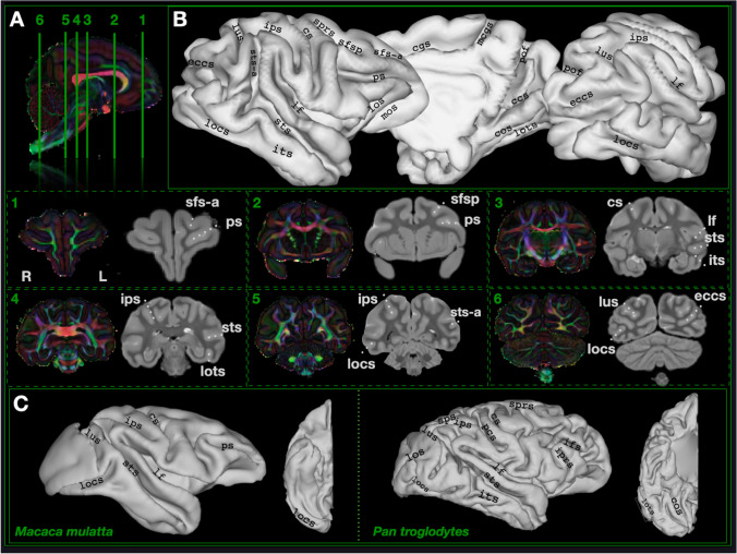
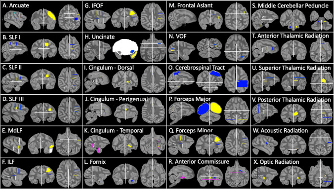
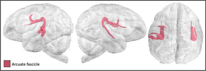
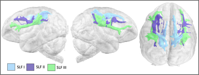
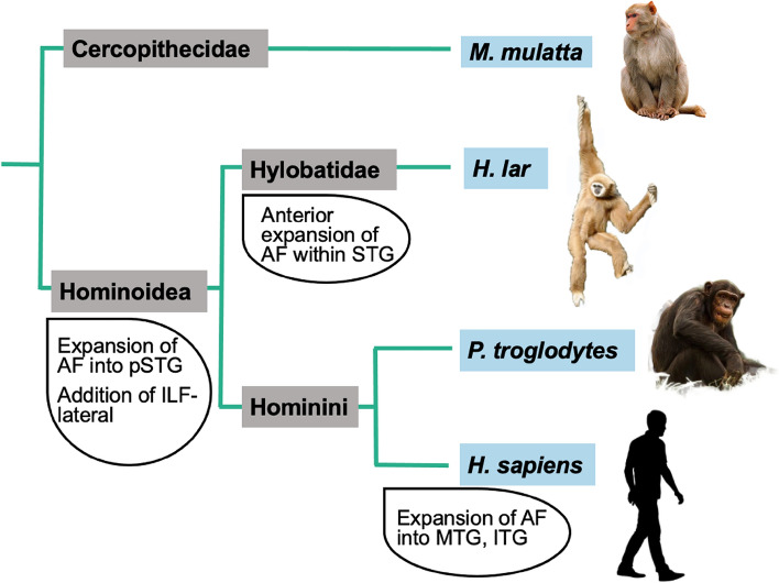
The recent development of methods for constructing directly comparable white matter atlases in primate brains from diffusion MRI allows us to probe specializations unique to humans, great apes, and other primate taxa. Here, we constructed the first white matter atlas of a lesser ape using an ex vivo diffusion-weighted scan of a brain from a young adult (5.5 years) male lar gibbon. We find that white matter architecture of the gibbon temporal lobe suggests specializations that are reminiscent of those previously reported for great apes, specifically, the expansion of the arcuate fasciculus and the inferior longitudinal fasciculus in the temporal lobe. Our findings suggest these white matter expansions into the temporal lobe were present in the last common ancestor to hominoids approximately 16 million years ago and were further modified in the great ape and human lineages. White matter atlases provide a useful resource for identifying neuroanatomical differences and similarities between humans and other primate species and provide insight into the evolutionary variation and stasis of brain organization.

Keywords: DWI; Evolution; Fasciculus; Hominoid; Tractography.


Conflict of interest statement

None to declare.

Figures

Fig. 1
Fig. 1
Sulcal boundaries in the lar gibbon with comparative anatomy in macaques and chimpanzees. A Coronal sections showing diffusion and grey matter at six locations throughout the brain. B Gibbon cortical surface reconstruction with sulcal labeling. C Macaque (three subject average from Roumazeilles et al. (2022)), gibbon, and chimpanzee (single subject from Roumazeilles et al. (2020)) for comparison. ccs calcarine sulcus, cgs cingulate sulcus, mcgs margin of the cingulate sulcus, cos collateral sulcus, cs central sulcus, eccs external calcarine sulcus, ifs inferior frontal sulcus, iprs inferior precentral sulcus, ips intraparietal sulcus, its inferior temporal sulcus, lus lunate sulcus, lf lateral fissure, locs lateral occipital sulcus, lots lateral occipitotemporal sulcus, mos medial orbital sulcus, sprs superior precentral sulcus, pof parieto-occipital fissure, ps principal sulcus, sfs-a superior frontal sulcus—anterior ramus, sfsp superior frontal sulcus—posterior ramus, sts superior temporal sulcus, sts-a superior temporal sulcus—ramus
Fig. 2
Fig. 2
Lar gibbon tractography recipes. Seed ROIs (yellow), target ROIs (blue), exclusion masks (white), stop masks (fuchsia). Left hemisphere protocols are displayed. Arcuate protocol uses two targets: target 1 (light blue); target 2 (dark blue)
Fig. 3
Fig. 3
Arcuate fascicle in the lar gibbon
Fig. 4
Fig. 4
SLFs I, II, and III in the lar gibbon
Fig. 5
Fig. 5
A Major fasciculi that course through the temporal lobe: (IFOF (inferior fronto-occipital fascicle), ILF (inferior longitudinal fascicle), MdLF (middle longitudinal fascicle) and UF (uncinate fascicle) in the lar gibbon. B Lateral (ILF-lat) and medial (ILF-med) subdivisions of the ILF in the gibbon
Fig. 6
Fig. 6
Cingulum bundle and fornix in the lar gibbon
Fig. 7
Fig. 7
AC (anterior commissure), FA (frontal aslant), VOF (vertical occipital fasciculus), and MCP (middle cerebellar peduncle) in the lar gibbon
Fig. 8
Fig. 8
Corticospinal and somatosensory pathways (CSP), forceps major (FMA) and minor (FMI) in the lar gibbon
Fig. 9
Fig. 9
Anterior, superior, and posterior thalamic radiations (ATR, STR, and PTR) in the lar gibbon
Fig. 10
Fig. 10
Optic and acoustic radiations in the lar gibbon
Fig. 11
Fig. 11
Summary of findings in evolutionary context

References

    1. Amiez C, Sallet J, Hopkins WD, Meguerditchian A, Hadj-Bouziane F, Ben Hamed S, Wilson CRE, Procyk E, Petrides M (2019) Sulcal organization in the medial frontal cortex provides insights into primate brain evolution. Nat Commun 10:1–14 - PMC - PubMed
    1. Apfelbach R (1972) Electrically elicited vocalizations in the Gibbon Hylobates lar (Hylobatidae), and their behavioral significance. Z Tierpsychol 30:420–430 - PubMed
    1. Avants BB, Tustison NJ, Song G, Cook PA, Klein A, Gee JC (2011) A reproducible evaluation of ANTs similarity metric performance in brain image registration. Neuroimage 54:2033–2044 - PMC - PubMed
    1. Azadbakht H, Parkes LM, Haroon HA, Augath M, Logothetis NK, de Crespigny A, D’Arceuil HE, Parker GJM (2015) Validation of high-resolution tractography against in vivo tracing in the macaque visual cortex. Cereb Cortex 25:4299–4309 - PMC - PubMed
    1. Behrens TEJ, Berg HJ, Jbabdi S, Rushworth MFS, Woolrich MW (2007) Probabilistic diffusion tractography with multiple fibre orientations: What can we gain? Neuroimage 34:144–155 - PMC - PubMed

MeSH terms

LinkOut - more resources