

Bilateral intracochlear schwannomas: histopathological confirmation and outcomes following tumour removal and cochlear implantation with lateral wall electrodes
- PMID: 37904024
- PMCID: PMC10663204
- DOI: 10.1007/s00106-023-01379-7
Bilateral intracochlear schwannomas: histopathological confirmation and outcomes following tumour removal and cochlear implantation with lateral wall electrodes
Abstract
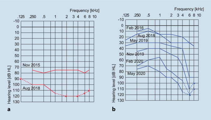
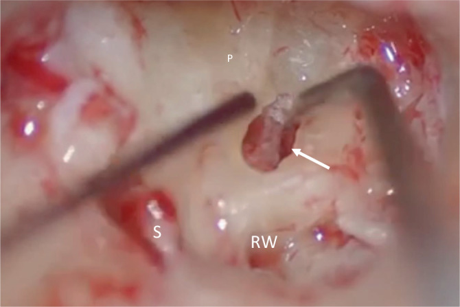
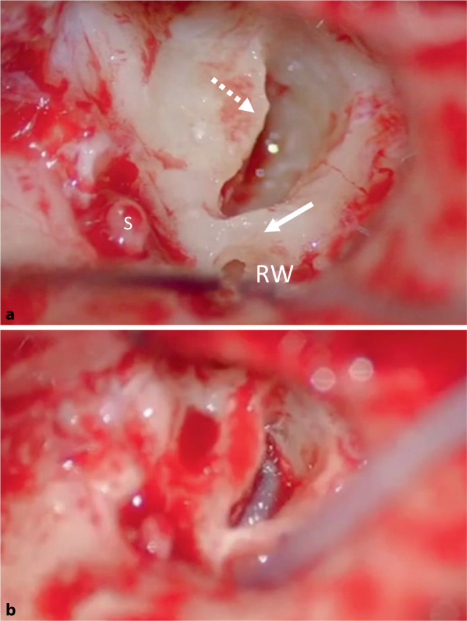
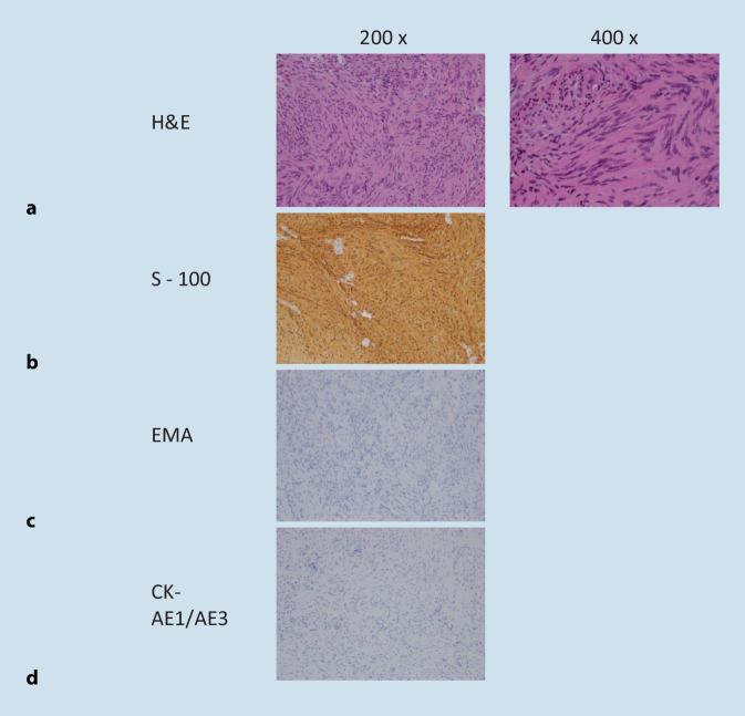
Intracochlear schwannomas (ICS) are very rare benign tumours of the inner ear. We present histopathological proof of the extremely rare bilateral occurrence of intracochlear schwannomas with negative blood genetic testing for neurofibromatosis type 2 (NF2). Bilateral schwannomas are typically associated with the condition NF2 and this case is presumed to have either mosaicism for NF2 or sporadic development of bilateral tumours. For progressive bilateral tumour growth and associated profound hearing loss, surgical intervention via partial cochleoectomy, tumour removal, preservation of the modiolus, and simultaneous cochlear implantation with lateral wall electrode carrier with basal double electrode contacts was performed. The right side was operated on first with a 14-month gap between each side. The hearing in aided speech recognition for consonant-nucleus-consonant (CNC) phonemes in quiet improved from 57% to 83% 12 months after bilateral cochlear implantation (CI). Bilateral intracochlear schwannomas in non-NF2 patients are extremely rare but should be considered in cases of progressive bilateral hearing loss. Successful tumour removal and cochlear implantation utilizing a lateral wall electrode is possible and can achieve good hearing outcomes.
Intracochleäre Schwannome (ICS) sind sehr seltene benigne Tumoren des Innenohrs. In der vorliegenden Arbeit wird die histopathologische Bestätigung des extrem seltenen bilateralen Vorkommens von intracochleären Schwannomen mit negativem genetischem Bluttest auf Neurofibromatose 2 (NF2) vorgestellt. Bilaterale Schwannome sind typischerweise mit NF2 assoziiert, und bei dem vorliegenden Fall wird vermutet, dass entweder ein Mosaik für NF2 oder die sporadische Entwicklung von bilateralen Tumoren besteht. Bei progressivem bilateralem Tumorwachstum und damit einhergehender hochgradiger Schwerhörigkeit wurde eine chirurgische Intervention mit partieller Cochleoektomie, Tumorentfernung, Erhalt des Modiolus und gleichzeitiger Cochleaimplantation unter Einsatz eines „lateral wall“-Elektrodenträgers mit basal doppelten Elektrodenkontakten durchgeführt. Zuerst wurde die rechte Seite operiert und mit einem Abstand von 14 Monaten die zweite Seite. Das Sprachverstehen für Konsonant-Nucleus-Konsonant(CNC)-Phoneme in Ruhe verbesserte sich 12 Monate nach bilateraler Cochleaimplantation von 57 auf 83%. Bilaterale intracochleäre Schwannome bei Nicht-NF2-Patienten sind extrem selten, aber sollten in Fällen mit progressiver bilateraler Schwerhörigkeit in Erwägung gezogen werden. Die erfolgreiche Tumorentfernung und Cochleaimplantation unter Verwendung von „lateral wall“-Elektrodenträgern ist möglich und kann zu guten Hörergebnissen führen.
Keywords: Acoustic neuroma; Cochlea; Cochlear implant; Inner ear; Intralabyrinthine.
© 2023. The Author(s).
Conflict of interest statement
M.E. Quick, S. Withers, S.K. Plontke, R. Chester-Browne and J. Kuthubutheen declare that they have no competing interests.
Figures


Similar articles
-
[Bilateral intracochlear schwannomas in a patient with no genetic or clinical features of neurofibromatosis type 2. German version].HNO. 2020 Jul;68(7):534-538. doi: 10.1007/s00106-019-00751-w. HNO. 2020. PMID: 31758201 German.
-
Cochlear Implantation in a Patient with Neurofibromatosis Type 2 and an Intracochlear Schwannoma: A Case Report and Literature Review.ORL J Otorhinolaryngol Relat Spec. 2022;84(5):425-428. doi: 10.1159/000522473. Epub 2022 May 10. ORL J Otorhinolaryngol Relat Spec. 2022. PMID: 35537404 Review.
-
Cochlear Implantation in Patients With Intracochlear and Intralabyrinthine Schwannomas.Otol Neurotol. 2016 Jul;37(6):647-53. doi: 10.1097/MAO.0000000000001016. Otol Neurotol. 2016. PMID: 27273407
-
Surgical Outcomes of Simultaneous Cochlear Implantation and Intracochlear Schwannoma Removal.Otolaryngol Head Neck Surg. 2023 Sep;169(3):660-668. doi: 10.1002/ohn.300. Epub 2023 Feb 17. Otolaryngol Head Neck Surg. 2023. PMID: 36807253
-
Ipsilateral Cochlear Implantation in the Presence of Observed and Irradiated Vestibular Schwannomas.Ann Otol Rhinol Laryngol. 2020 Dec;129(12):1229-1238. doi: 10.1177/0003489420935482. Epub 2020 Jun 18. Ann Otol Rhinol Laryngol. 2020. PMID: 32551844 Review.
Cited by
-
Cochlear implantation in patients with inner ear schwannomas: a systematic review and meta-analysis of audiological outcomes.Eur Arch Otorhinolaryngol. 2024 Dec;281(12):6175-6186. doi: 10.1007/s00405-024-08818-3. Epub 2024 Jul 11. Eur Arch Otorhinolaryngol. 2024. PMID: 38992191 Free PMC article.
References
-
- Dubernard X, Somers T, Veros K, Vincent C, Franco-Vidal V, Deguine O, et al. Clinical presentation of intralabyrinthine schwannomas: a multicenter study of 110 cases. Otol Neurotol Off Publ Am Otol Soc Am Neurotol Soc [and] Eur Acad Otol Neurotol. 2014;35(9):1641–1649. doi: 10.1097/MAO.0000000000000415. - DOI - PubMed
-
- Kennedy RJ, Shelton C, Salzman KL, Davidson HC, Harnsberger HR. Intralabyrinthine schwannomas: diagnosis, management, and a new classification system. Otol Neurotol Off Publ Am Otol Soc Am Neurotol Soc [and] Eur Acad Otol Neurotol. 2004;25(2):160–167. doi: 10.1097/00129492-200403000-00014. - DOI - PubMed
MeSH terms
LinkOut - more resources
Full Text Sources
Medical
Research Materials
Miscellaneous
