. 2023 Nov;4(11):1592-1609.
doi: 10.1038/s43018-023-00652-6. Epub 2023 Oct 30.

Structural surfaceomics reveals an AML-specific conformation of integrin β2 as a CAR T cellular therapy target

Affiliations

Structural surfaceomics reveals an AML-specific conformation of integrin β2 as a CAR T cellular therapy target

Kamal Mandal et al. Nat Cancer. 2023 Nov.

Abstract

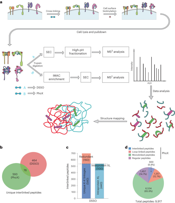
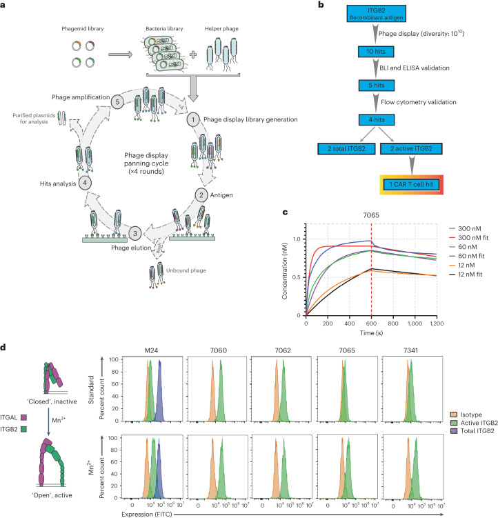
Safely expanding indications for cellular therapies has been challenging given a lack of highly cancer-specific surface markers. Here we explore the hypothesis that tumor cells express cancer-specific surface protein conformations that are invisible to standard target discovery pipelines evaluating gene or protein expression, and these conformations can be identified and immunotherapeutically targeted. We term this strategy integrating cross-linking mass spectrometry with glycoprotein surface capture 'structural surfaceomics'. As a proof of principle, we apply this technology to acute myeloid leukemia (AML), a hematologic malignancy with dismal outcomes and no known optimal immunotherapy target. We identify the activated conformation of integrin β2 as a structurally defined, widely expressed AML-specific target. We develop and characterize recombinant antibodies to this protein conformation and show that chimeric antigen receptor T cells eliminate AML cells and patient-derived xenografts without notable toxicity toward normal hematopoietic cells. Our findings validate an AML conformation-specific target antigen and demonstrate a tool kit for applying these strategies more broadly.


Conflict of interest statement

K.M., J.J.A., S.S.S. and A.P.W. have filed a patent related to the antibody sequences described herein. A.P.W. has received research funding from Genentech. C.S. has received research funding from Revolution Medicines, Abbvie and Erasca and has served on advisory boards for Genentech, Abbvie and Astellas. C.M.R. has equity in Allovir and Marker Therapeutics, has received research support from Tessa Therapeutics and is on the Scientific Advisory Board of Tessa Therapeutics and Marker. The spouse of C.M.R. has served on advisory boards for Walking Fish Therapeutics, CellGenix, Marker Therapeutics, Tessa Therapeutics, Abintus, Allogene, Bellicum Pharmaceuticals, Bluebird Bio, Athenex, Memgen, Turnstone Biologics, Coya Therapeutics, TScan Therapeutics, Onkimmune, Poseida Therapeutics and Allovir. The other authors declare no competing interests.

Figures

Fig. 1
Fig. 1. XL–MS and surface glycoprotein capture strategy to identify conformation-specific cancer antigens.
a, Schematic flow diagram of the ‘structural surfaceomics’ approach. b, Venn diagram showing the total number of cross-linked peptides identified from the two different approaches (tandem MS (MS2) and multistage MS (MS3) based). PhoX and DSSO were used as cross-linkers for the MS2 and MS3 approaches, respectively. c, Bar graph showing the distribution of inter- and intraprotein cross-links (XL) from MS3-based (DSSO) XL–MS. d, Pie chart showing the distribution of the various types of cross-links obtained from PhoX MS2-based XL–MS. All cross-links were identified with a ≤1% FDR (see Methods for details). ‘Regular’ peptides indicate that no PhoX modification was detected on any lysines.
Fig. 2
Fig. 2. Activated integrin β2 is a conformationally selective antigen in AML.
a, Identified cross-linked peptides mapped on to the crystal structure of the integrin αL/integrin β2 heterodimer (PDB 5E6R). b, Flow cytometry histogram plot showing expression of total and activated integrin β2 on AML (top) and B cell lines (bottom; BV-173 and Namalwa). The y axis represents percent count normalized to mode. The gating strategy is shown in Supplementary Information 1a. Data are representative of n = 4 (Nomo1), 2 (THP-1) and 1 (all others) independent experiments. c, Flow cytometry plot showing the absence of active integrin β2 on CD34+ HSPCs from GM-CSF-mobilized peripheral blood. The gating strategy is shown in Supplementary Information 1b. Deidentified human samples were used for this analysis (n = 5 independent donors). d, Representative flow cytometry histogram plots showing the expression of active integrin β2 on primary AML cells. The y axis represents percent count normalized to mode. The gating strategy is shown in Supplementary Information 1c. Data are representative of n = 10 total deidentified samples. e, Heat map showing inverse expression patterns of ITGB2 against other AML targets in publicly available primary AML RNA-seq data. The color bar represents maximum expression in each row based on normalized read counts. The sample sizes of BEAT AML (adult), TARGET (pediatric) and TCGA were 510, 255 and 150, respectively. f, Aggregated single-cell RNA-seq data showing essentially exclusive expression of ITGB2 in hematopoietic tissue; data were obtained from the Human Protein Atlas; nTPM, normalized transcripts per million.
Fig. 3
Fig. 3. Antibody 7065 binds preferentially to the active conformation of integrin β2.
a, Schematic flow diagram of the phage display selection strategy used for developing antibodies to integrin β2. b, Schematic flow diagram showing triage of antibodies obtained from the phage display library and the downstream validation/funneling to identify an active integrin β2 binder. c, Representative BLI plot showing determination of binding affinity (Kd) of the 7065 antibody to integrin αL/integrin β2; n = 3 different concentrations of antibody were used for this experiment (see also Extended Data Fig. 6c). d, Flow cytometry analysis of Jurkat T-ALL cells in the presence and absence of 2 mM Mn2+ ions to determine/identify antibodies with specificity to active integrin β2. The y axis represents percent count normalized to mode. The gating strategy is shown in Supplementary Information 1a. Data are representative of n = 2 independent experiments.
Fig. 4
Fig. 4. aITGB2 CAR T cells derived from the 7065 antibody are cytotoxic to AML cells.
a, Schematic diagram of the CAR T construct used; TM, transmembrane; costim, co-stimulatory domain. b, Luciferase-based cytotoxicity of the aITGB2 CAR T design in Nomo1 and THP-1 AML cell lines. Data are representative of n = 3 independent experiments with similar results. Each experiment was performed in triplicate. c, Incucyte live-cell imaging data demonstrating efficient cytotoxicity of aITGB2 CAR T cells against Nomo1 cells at two different E:T ratios (1:1 and 1:10) over a 5-d period. CAR T cells were labeled with GFP, and tumor cells (Nomo1) were labeled with mCherry to facilitate fluorescence-based quantification. The y axis represents integrated fluorescence used as a proxy to monitor cell proliferation. Data are from a single experiment performed with six replicates. CU, calibrated units. d, Flow cytometry histogram showing a successfully generated ITGB2-knockout version of Nomo1 cells using CRISPR–Cas9. The y axis represents percent count normalized to mode. The gating strategy is shown in Supplementary Information 1a; KO, knockout; WT, wild-type. e, Luciferase-based cytotoxicity data showing specific activity of aITGB2 CAR T cells against wild-type Nomo1 cells and not against ITGB2-knockout Nomo1 cells (the E:T ratio was 1:1 with overnight incubation). Data are representative of n = 2 independent experiments with similar results. Each experiment was performed in triplicate. The luciferase signals of the cytotoxicity assays were normalized against untransduced CAR T cells of their respective E:T ratios. Only aITGB2 CAR T cell manufacturing involved knocking out ITGB2.
Fig. 5
Fig. 5. Toxicity assessment of aITGB2 CAR T cells demonstrates a promising safety profile.
a, Flow cytometry-based cytotoxicity assay showing specificity of aITGB2 CAR T cells to activated peripheral blood T cells that harbor activated integrin β2 (lower right quadrant with CAR and CD3+ T cells). Both resting and activated conditions were performed in overnight coculture assays with aITGB2 CAR T cells. The gating strategy is similar to that shown in Supplementary Information 1b. b, Flow cytometry analysis showing successful activation of T cells and partial abundance of activated integrin β2 on activated T cells. The gating strategy is similar to that shown in Supplementary Information 1b. c, Quantitative analysis of active T cell depletion data in a. Data are representative of n = 2 independent experiments with similar results, one of which was performed in triplicate. d, Clonogenic assay showing no impact of aITGB2 CAR T cells against CD34+ HSPCs from GM-CSF-mobilized peripheral blood. The E:T ratio was 1:1 (see Methods section for details). Data are representative of n = 2 independent experiments with similar results. Each experiment was performed in triplicate; c.f.u., colony-forming units; GEMM, granulocyte, erythrocyte, monocyte, megakaryocyte; GM, granulocyte, monocyte; G, granulocyte; M, monocyte; E, erythrocyte; b.f.u., burst-forming units. e, Flow cytometry analysis showing no discernible impact of aITGB2 CAR T cells against T cells and B cells. The y axis represents percent count normalized to mode. The gating strategy is similar to that shown in Supplementary Information 1b. Also see Extended Data Fig. 9d. f, Schematic flow diagram for the generation of HIS mice. g, Representative flow cytometry data from HIS mice showing apparent non-toxicity of aITGB2 CAR T cells against myeloid cells (CD14+). All events were used for gating and analysis. Data are representative of n = 4 mice and 6 d after CAR T cell treatment. h, Quantification of hCD45+ data in g; n = 4 mice. The gating strategy is similar to that shown in Supplementary Information 2e. P values were calculated by two-tailed t-test (P = 0.0008 (aITGB2 CAR versus CD33 CAR) and P = 0.0007 (empty CAR versus CD33 CAR)); ***P ≤ 0.001; NS, not significant. i, Complete blood count (CBC) profiling of HIS mice treated with aITGB2 CAR T cells on day 5 (data are from n = 4 mice). Only aITGB2 CAR T cell manufacturing involved knocking out ITGB2. For all the in vitro cytotoxicity assays, the E:T ratio was 1:1 with overnight incubation. All mice used were females; RBC, red blood cells.
Fig. 6
Fig. 6. Efficacy of aITGB2 CAR T cells against AML models in vivo.
a, Survival of NSG mice implanted with two independent AML PDX models and treated with aITGB2, anti-CD33 or empty CAR T cells; n = 6 mice per arm. In total, 2 × 106 AML tumor cells were injected on day 0, and 5 × 106 CAR T cells were injected on day 5. P values were determined by log-rank test (P = 0.009 (ITGB2 CAR T cells versus empty CAR T cells, PDX-A); P = 0.0068 (ITGB2 CAR T cells versus empty CAR T cells, PDX-B); P = 0.044 (CD33 CAR T cells versus ITGB2 CAR T cells, PDX-B)); **P ≤ 0.01; *P ≤ 0.05. b, Flow cytometry histogram plots of peripheral blood draws showing tumor burden at 8 weeks after tumor injection for PDX-A and at 3.5 weeks for PDX-B (also see Extended Data Fig. 10a,b). Naive control mice have no human cells (AML tumor or CAR T cells) injected and were used to assess background noise in the flow cytometry assay. The y axis represents percent count normalized to mode. The gating strategy is similar to that shown in Supplementary Information 2e. Data are representative of n = 6 mice per arm. Plots of only live animals are provided at the respective time points. c, Spleen ultrasonography (USG) from animals treated with empty CAR T cells compared to animals treated with CD33 or aITGB2 CAR T cells. All mice alive at day 49 after tumor implantation were scanned. Data are from mice still surviving at this time; n = 5 (aITGB2), n = 6 (CD33) and n = 3 (empty). d, BLI imaging showing the efficacy of aITGB2 CAR T cells against the intravenously implanted AML cell line Nomo1 (n = 6 mice per arm). e, Quantitative analysis of bioluminescence intensity of the mice in d plotted individually (n = 6 mice). A two-tailed Mann–Whitney test was used for statistical analysis of bioluminescence quantification (P = 0.0087 (day 33) and P = 0.0022 (day 41) ITGB2 CAR T cells versus empty CAR T cells); **P ≤ 0.01. Only aITGB2 CAR T cell manufacturing involved knocking out ITGB2. All statistical data are represented as mean ± s.e.m. All mice used in ac were females, and those in d–e were male.
Extended Data Fig. 1
Extended Data Fig. 1. Ving and XL-MS SEC.
a, Schematic workflow describing the working principle of Ving. b, Representative SEC trace of peptides obtained from DSSO cross-linked samples. c, Representative SEC trace of peptides obtained from PhoX cross-linked samples. For both strategies (DSSO and PhoX), samples were processed in 4 separate batches and each time the SEC trace pattern was similar. XL peptides refers to cross-linked peptides.
Extended Data Fig. 2
Extended Data Fig. 2. Representative MS spectra demonstrating the MS3 based strategy of XL-MS.
Example MS spectra demonstrating the MS3 based strategy of XL-MS. The cross-linked peptides of 811.57 m/z is selected for MS2, at which step the cross-linker is cleaved in the collision cell generating two separate peptides of 606.26 m/z and 942.45 m/z, respectively. These two high-abundance peptides are then selected for MS3 where they undergoes full fragmentation for peptide identification. We also note the respective modification on Lysine residues resulting from the cross-linker.
Extended Data Fig. 3
Extended Data Fig. 3. Discerning active integrin β2 expression.
a, Cartoon diagram showing proposed inactive and active conformations of ITGB2. b, Flow cytometry plot showing presence of total integrin β2 on CD34+ HSPCs from GM-CSF mobilized peripheral blood. Cells were gated on singlet cells for analysis. Deidentified patient samples were used for this analysis (n = 5, independent donors). c, Flow cytometry analysis showing expression of active ITGB2 in PDX models of AML (PDX-A and PDX-B). The y-axis represents percent count normalized to mode. Cells were gated on human CD45+ population cells for analysis. Representative plot from n = 2 separate PDX models of AML.
Extended Data Fig. 4
Extended Data Fig. 4. ITGB2 transcript expression.
a, AML subtype specific expression analysis of ITGB2 and other notable AML targets of patient samples from TARGET database. b, Single cell sequencing data showing expression of notable AML target across normal human tissues and immune cells (adapted from Human Protein Atlas).
Extended Data Fig. 5
Extended Data Fig. 5. Initial anti-active integrin β2 CAR-T designs.
Luciferase based cytotoxicity analysis of M24 and A57 antibody derived CAR-T cells against AML cell line Nomo-1 (Data from single experiment performed in triplicate). The luciferase signals of the cytotoxicity assays were normalized against untransduced CAR-T of their respective E:T ratios.
Extended Data Fig. 6
Extended Data Fig. 6. Characterizing recombinant antibodies to integrin β2.
a, Representative SEC trace of the antibody 7065 showing distinct peak, for quality check. b. Non-specific ELISA panel showing specificity profiles of the antibodies obtained from phage display selection. (Data from single experiment performed in duplicates) c, Representative BLI plots showing binding affinities (KD) of the antibodies against ITGB2 with their alpha partners. Each experiment was performed with n = 3 different concentrations of antibody. d, Flow cytometry analysis showing binding of antibody 7065 on the AML cells. The y-axis represents percent count normalized to mode. Gating strategy shown in Supplementary Information 1a. n = 3 independent experiments. e, Flow cytometry analysis showing binding of antibody 7065 on the primary AML cells, gated on CD34+ blasts. The y-axis represents percent count normalized to mode. Gating strategy shown in Supplementary Information 1c. n = 7 independent patient samples.
Extended Data Fig. 7
Extended Data Fig. 7. Evaluation of aITGB2 CAR-T designs incorporating recombinant antibodies.
a, Flow cytometry screen for cytotoxicity and activation status of aITGB2 CAR-T designs vs. AML cell line Nomo1 (n = 1 for each design). Similarly, as a demonstration of specificity, cytotoxicity and activation status was also checked against AMO-1 (multiple myeloma cell line that does not express integrin β2). Cells were gated on single cells for analysis. Gating strategy shown in Supplementary Information 2a. b, Flow cytometry analysis showing absence of ITGB2 in AMO-1. Cells were gated on single cells for analysis. Flow cytometry gating strategy similar to shown in Supplementary Information 1a. c, Flow cytometry analysis showing knockout efficiency of the various sgRNA used for knocking out ITGB2 in primary T cells (n = 1 for each sgRNA). Cells were gated on single cells for analysis. Flow cytometry gating strategy similar to that shown in Supplementary Information 1a. d, Plot showing proliferation of aITGB2-CAR-T cells, ‘with ITGB2 knockout’ vs ‘Non-Targeting (NT) – control’. Scrambled sgRNA was used for NT-control. (Data from single experiment performed in triplicate). e, Plots showing off-target analysis of the sgRNA used for CAR-T manufacturing using CRISPOR tool. Top 5 potential hits were assessed. f, Degranulation assay of aITGB2 and anti-CD33 CAR-T against Nomo1 based on CD107a staining. CAR positivity denoted by GFP tag on CAR construct. E:T ratio was 1:1 and 6 hours incubation time (n = 1). Cells were gated on single cells for analysis. Gating strategy similar to shown in Supplementary Information 2a. g, Luciferase assay-based cytotoxicity analysis showing cytotoxicity of aITGB2-CAR-T cells against AML cell lines with varying antigen density (Representative of n = 2 independent experiments with similar results, each experiment performed in triplicate). The flow cytometry data showing antigen density has been repurposed from Fig. 2b. Only aITGB2 CAR-T manufacturing involved knocking out integrin β2, and only in (f) and (g).
Extended Data Fig. 8
Extended Data Fig. 8. Additional aITGB2 CAR-T evaluation.
a, Luciferase assay-based cytotoxicity analysis showing efficacy of 7065 based aITGB2 CAR-T with 1x – 4x Gly4Ser (L1-L4) linker between heavy and light chain (Data from single experiment performed in triplicate). The luciferase signals of the cytotoxicity assays were normalized against untransduced T-cells of their respective E:T ratios. b, Bar plots showing cytotoxicity of aITGB2 CAR-T against primary AML patient samples. n = 3 technical replicates and 3 independent patient samples. E:T ratio was 3:1 with overnight incubation. Cells were gated on CD34+ cells for analysis. Gating strategy similar to Supplementary Information 1c. c, Cytokine profiling of aITGB2 CAR-T comparing against Empty CAR-T control upon pre vs post tumor exposure at E:T ratio of 1:1 for overnight incubation (Data from single experiment performed in triplicate). df, Representative flow cytometry-based histogram analysis showing activation markers (d), exhaustion markers (e) and memory markers (f) of CAR-T in pre vs post tumor exposure at E:T ratio of 1:1 for overnight incubation. Gating strategy similar to Supplementary Information 2d. Antibody dilutions used in df was 1:50. Only aITGB2 CAR-T manufacturing involved knocking out integrin β2. All the statistical data in this figure are represented as mean ± SEM.
Extended Data Fig. 9
Extended Data Fig. 9. Determining specificity of aITGB2 CAR-T.
a, Luciferase assay-based cytotoxicity analysis showing no activity of aITGB2 CAR-T vs. Namalwa (B-ALL) line which does not harbor active ITGB2 although it does have total form of ITGB2 (see Fig. 2b). Nomo-1 as the positive control (Data from single experiment performed in triplicate). The luciferase signals of the cytotoxicity assays were normalized against untransduced T-cells of their respective E:T ratios. b, Flow cytometry analysis showing absence of active ITGB2 and presence of total ITGB2 in T and B cells (n = 3 independent experiments). Cells were gated on single cells for analysis. Gating strategy similar to that shown in Supplementary information 1b. c, Flow cytometry analysis showing no discernible impact of aITGB2 CAR-T against CD34+ HSPCs from GM-CSF mobilized peripheral blood. The y-axis represents percent count normalized to mode. Gating strategy similar to that shown in Supplementary information 2b (n = 1 donor). d, Flow cytometry analysis showing non-specific depletion of B cells with aITGB2 and anti-CD33 CAR-T (Representative of n = 2 independent experiments with similar results, each experiment performed in triplicate). e, Flow cytometry plots showing activation of pathogen-specific T cells using CD69 as a marker. f, Quantitative analysis showing no depletion of pathogen-specific T cells with aITGB2 CAR-T when at resting state, but moderate depletion when activated exogenously. Gating strategy shown in Supplementary information 2f (Data from single experiment performed in triplicate). g, Flow cytometry analysis showing presence of active ITGB2 in myeloid cells (n = 3 independent experiments). Cells were gated on single cells for analysis. Gating strategy similar to that shown in Supplementary information 2c. h, Flow cytometry analysis showing cytotoxicity of aITGB2-CAR against neutrophils and monocytes in vitro. Cells were gated on single cells for analysis. i, Quantitative plot showing absence of cytotoxicity of aITGB2 CAR-T against human CD45+ cells in HIS mice at noted time points. Peripheral blood samples were used for this analysis. CD33 CAR-T serves as a positive control where significant cytotoxicity was observed (n = 5 mice in each arm). Two-tailed t-test were performed for statistical analysis. [p-value = 0.009 (2-week, ITGB2-CAR vs CD33-CAR); p-value = 0.047 (2-week, Empty-CAR vs CD33-CAR); p-value = 0.035 (4-week, ITGB2-CAR vs CD33-CAR)]; *p ≤ 0.05; **p ≤ 0.01 j, Flow cytometry analysis showing cross reactivity of 7065 antibody against the murine ITGB2 on S49.1 cell line. The y-axis represents percent count normalized to mode. Cells were gated on single cells for analysis. Gating strategy similar to shown in Supplementary information 1a. k, Quantitative bar plots showing validation of mice aITGB2 CAR-T for its toxicity against murine S49.1 cells. E:T ratio was 2:1 with overnight coculture. hCD19 CAR-T, being specific for human CD19 only, was used as a negative control in murine CAR-T format. Gating strategy shown in Supplementary information 2f. (Data from single experiment performed in triplicate). l, Flow cytometry analysis showing knockout efficiency of the mice sgRNA used for knocking out ITGB2 in primary mice T cells (n = 1). Cells were gated on single cells for analysis. Flow cytometry gating strategy similar to that shown in Supplementary Information 1a. m, Complete blood count profiling of HIS mice treated with aITGB2 CAR-T periodically over 6 weeks (data from n = 6 mice). Only aITGB2 CAR-T manufacturing involved knocking out integrin β2. All the statistical data in this figure are represented as mean ± SEM. All the mice used in this figure were females.
Extended Data Fig. 10
Extended Data Fig. 10. aITGB2 CAR-T efficacy in PDX models.
a, Flow cytometry analysis and bar graph of peripheral blood draw showing tumor burden at 6 and 8 weeks post tumor injection of PDX-A. The y-axis represents percent count normalized to mode. Cells were gated on single cells for analysis. Representative of data from n = 6 mice per arm; plots of only mice alive at designated time point. b, Flow cytometry analysis and bar graph of peripheral blood draw showing tumor burden at 3.5 weeks post tumor injection of PDX-B. The y-axis represents percent count normalized to mode. Cells were gated on single cells for analysis. Representative of data from n = 6 mice per arm; plots of only mice alive at designated time point. c, Plots showing expansion and persistence of aITGB2 CAR-T in mice peripheral blood, analyzed periodically for 3 months (n = 6 mice per arm, subsequent data points were from mice alive at that time point). d, Flow cytometry analysis showing active ITGB2 density of tumor cells harvested from relapse Nomo-1 mice model (n = 4 mice per condition). The y-axis represents percent count normalized to mode. Cells were gated on human CD45+ cells for analysis. Gating strategy similar to that shown in Supplementary information 2e. Only aITGB2 CAR-T manufacturing involved knocking out integrin β2. All the statistical data in this figure are represented as mean ± SEM. All the mice used in this figure were females.

References

    1. Lim WA, June CH. The principles of engineering immune cells to treat cancer. Cell. 2017;168:724–740. - PMC - PubMed
    1. Brooks JD. Translational genomics: the challenge of developing cancer biomarkers. Genome Res. 2012;22:183–187. - PMC - PubMed
    1. Beck A, Goetsch L, Dumontet C, Corvaïa N. Strategies and challenges for the next generation of antibody–drug conjugates. Nat. Rev. Drug Discov. 2017;16:315–337. - PubMed
    1. Brudno, J. N. & Kochenderfer, J. N. Toxicities of chimeric antigen receptor T cells: recognition and management. Blood127, 3321–3330 (2016). - PMC - PubMed
    1. Hosen N, et al. The activated conformation of integrin β7 is a novel multiple myeloma-specific target for CAR T cell therapy. Nat. Med. 2017;23:1436–1443. - PubMed

Publication types

MeSH terms