[Preprint]. 2023 Nov 2:2023.10.20.563371.
doi: 10.1101/2023.10.20.563371.

Exploiting the Cullin E3 Ligase Adaptor Protein SKP1 for Targeted Protein Degradation

Affiliations

Exploiting the Cullin E3 Ligase Adaptor Protein SKP1 for Targeted Protein Degradation

Seong Ho Hong et al. bioRxiv. .

Update in

Abstract

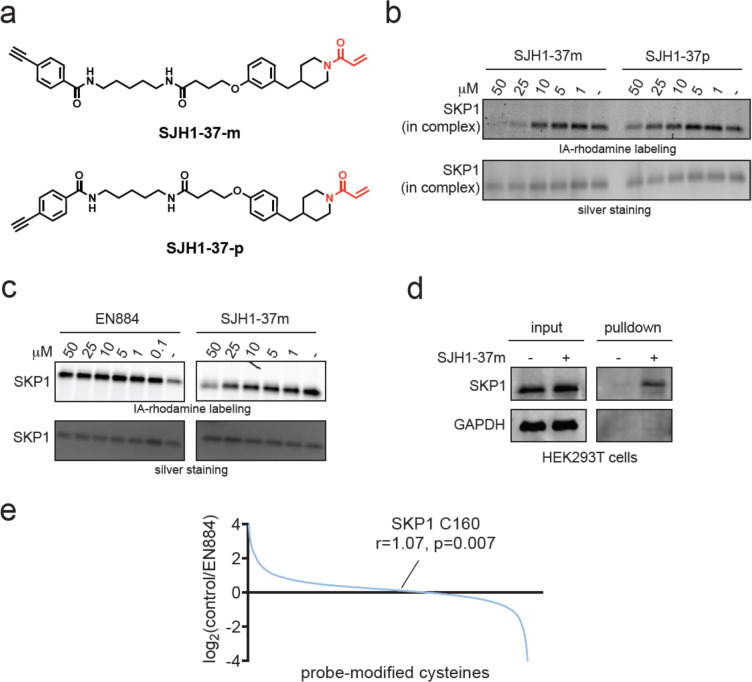
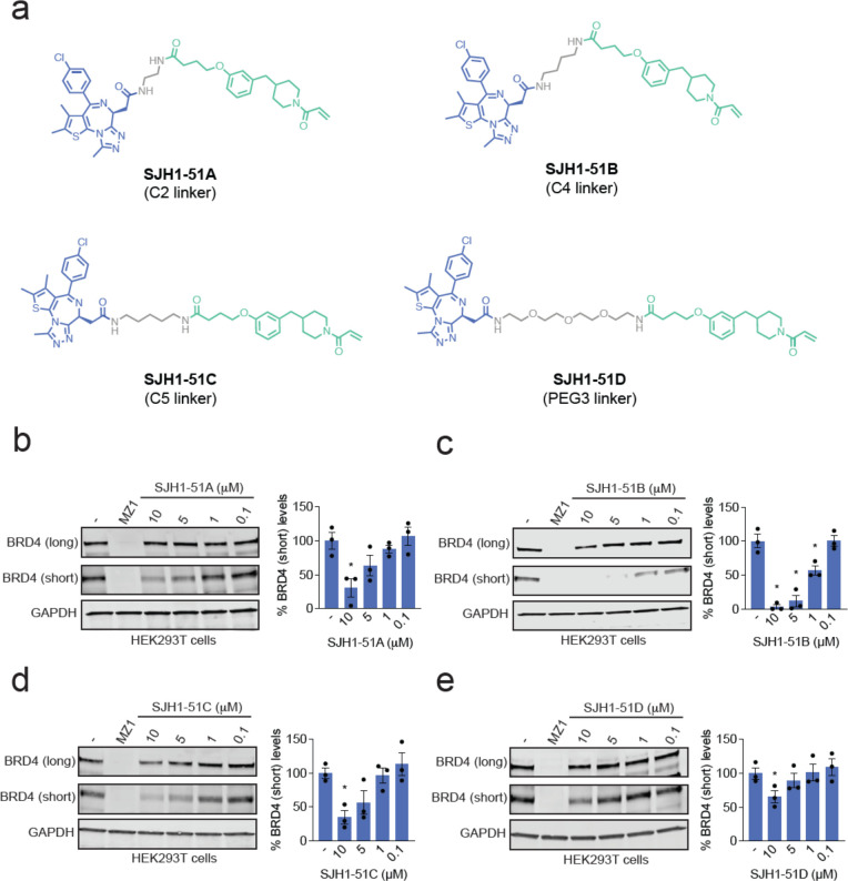
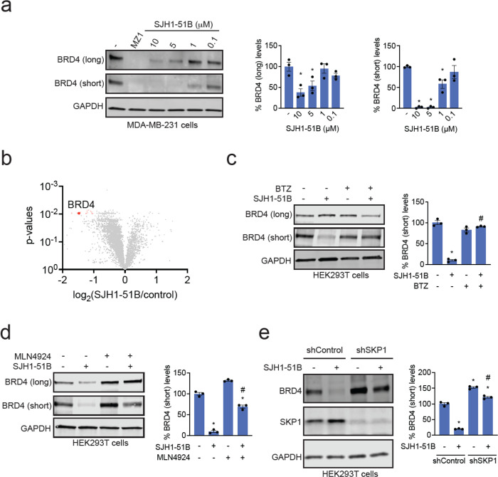
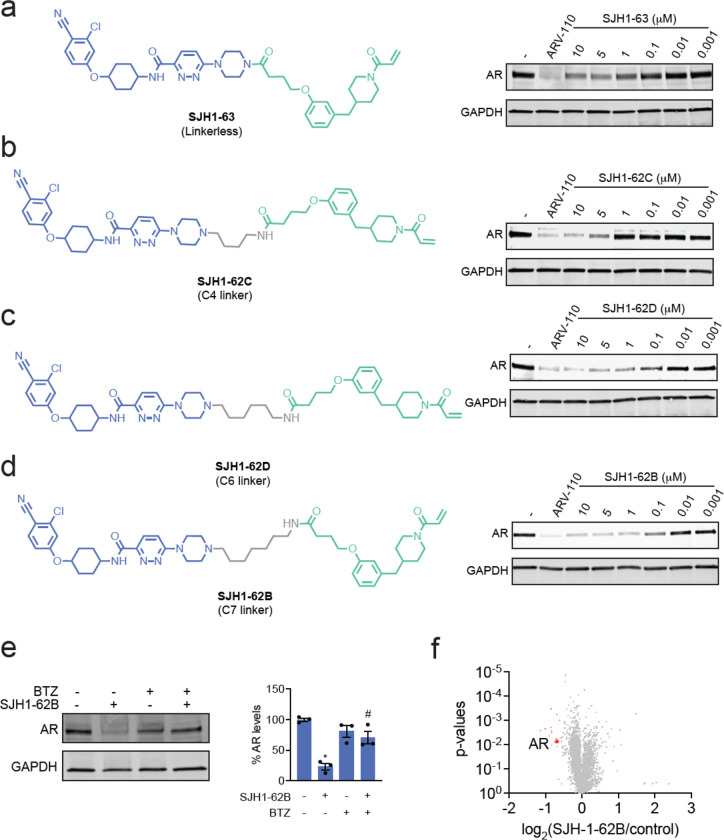
Targeted protein degradation with Proteolysis Targeting Chimeras (PROTACs) is a powerful therapeutic modality for eliminating disease-causing proteins through targeted ubiquitination and proteasome-mediated degradation. Most PROTACs have exploited substrate receptors of Cullin-RING E3 ubiquitin ligases such as cereblon and VHL. Whether core, shared, and essential components of the Cullin-RING E3 ubiquitin ligase complex can be used for PROTAC applications remains less explored. Here, we discovered a cysteine-reactive covalent recruiter EN884 against the SKP1 adapter protein of the SKP1-CUL1-F-box containing SCF complex. We further showed that this recruiter can be used in PROTAC applications to degrade neo-substrate proteins such as BRD4 and the androgen receptor in a SKP1- and proteasome-dependent manner. Our studies demonstrate that core and essential adapter proteins within the Cullin-RING E3 ubiquitin ligase complex can be exploited for targeted protein degradation applications and that covalent chemoproteomic strategies can enable recruiter discovery against these targets.

Keywords: AR; BRD4; CUL1; SKP1; activity-based protein profiling; androgen receptor; chemoproteomics; covalent; targeted protein degradation.


Conflict of interest statement

Competing Financial Interests Statement OWH was an employee of Bristol Myers Squibb when this study was initiated, but is now an employee of Lyterian Therapeutics. IEW was an employee of Bristol Myers Squibb when this study was initiated but is now a co-founder and the CEO of Lyterian Therapeutics. IEW is on the Scientific Advisory Boards of PAIVBio and Firefly Biologics. DKN is a co-founder, shareholder, and scientific advisory board member for Frontier Medicines and Vicinitas Therapeutics. DKN is a member of the board of directors for Vicinitas Therapeutics. DKN is also on the scientific advisory board of The Mark Foundation for Cancer Research, Photys Therapeutics, and Apertor Pharmaceuticals. DKN is also an Investment Advisory Partner for a16z Bio, an Advisory Board member for Droia Ventures, and an iPartner for The Column Group.

Figures

Figure 1.
Figure 1.. Discovery of a covalent SKP1 recruiter.
(a) Gel-based ABPP screening of a cysteine-reactive covalent ligand library (50 μM) against a rhodamine-functionalized iodoacetamide (IA-rhodamine) labeling of pure SKP1 protein in the SKP1-FBXO7-CUL1-RBX1 complex. Individual compound values are noted as percent of labeling compared to that of DMSO vehicle control. EN884 was the top hit that showed the most inhibition of probe labeling. (b) Structure of EN884 with the cysteine reactive acrylamide warhead in red. (c) gel-based ABPP of EN884 competition against IA-rhodamine labeling. Pure SKP1 in the SKP1-FBXO7-CUL1-RBX1 complex was pre-incubated with EN884 1 h prior to labeling with IA-rhodamine (100 nM) for 30 min after which proteins were separated by SDS/PAGE and visualized by in-gel fluorescence. Protein loading was assessed by silver staining. (d) Mass spectrometry analysis of EN884 modification on SKP1. The SKP1-FBXO7-CUL1-RBX1 complex was incubated with EN884 (50 μM) for 1 h and tryptic digests from the complex were subsequently analyzed for the EN884 covalent adduct on a cysteine. Shown is the MS/MS data for EN884 modification on SKP1 C160. (e) Shown is the location in the circle of SKP1 C160 in the CUL1 complex with SKP1 in blue, in complex with SKP2, RBX1, and CUL1. Shown is an SCF complex model derived from superimposing the crystal structures of Skp1-Skp2-Cks1 with p27 peptide (PDB ID: 2AST) and Cul1-Rbx1-Skp1-Skp2 (PDB ID: 1LDK). Data shown in (b) are from n=3 biologically independent replicates per group.
Figure 2.
Figure 2.. Characterization of covalent SKP1 recruiter.
(a) Structures of two alkyne-functionalized probes based on EN884. (b) Gel-based ABPP of both probes against IA-rhodamine labeling of pure SKP1 in the SKP1-FBXO7-CUL1-RBX1 complex as described in (a). (c) Gel-based ABPP of EN884 and SJH1-37m against pure SKP1 not in complex assessed as described in (b). (d) SKP1 pulldown using SJH1-37m probe. HEK293T cells were treated with DMSO vehicle or SJH1-37m (50 μM) for 4 h. Lysates were subjected to copper-catalyzed azide-alkyne cycloaddition (CuAAC) to append on a biotin enrichment handle after which probe-modified proteins were avidin-enriched and separated on SDS/PAGE and SKP1 and an unrelated protein GAPDH were detected by Western blotting. (e) isoDTB-ABPP analysis of EN884 in HEK293T cells. HEK293T cells were treated with DMSO vehicle or EN884 (50 μM) for 4 h, after which lysates were labeled with an alkyne-functionalized iodoacetamide probe (IA-alkyne) and control and treated cells were subjected to CuAAC with a desthiobiotin-azide with an isotopically light or heavy handle, respectively. After the isoDTB-ABPP procedure, probe-modified peptides were analyzed by LC-MS/MS and control vs treated probe-modified peptide ratios were quantified. Data shown in (b, c, d, e) are from n=3 biologically independent replicates per group.
Figure 3.
Figure 3.. SKP1-based BRD4 degraders.
(a) Structures of 4 BRD4 degraders using covalent SKP1 recruiters in green linked via a linker in grey to the BET family inhibitor JQ1 in blue via a C2, C4, or C5 alkyl linker or PEG3 linker. (b-e) BRD4 degradation in cells. HEK293T cells were treated with DMSO vehicle, MZ1 (1 μM), or each degrader for 24 h. The long and short isoforms of BRD4 and loading control GAPDH were assessed by Western blotting and bands were quantified by densitometry and normalized to GAPDH. Blots are representative of n=3 biologically independent replicates/group. Bar graphs show average ± sem and individual replicate values of the BRD4 short isoform. Significance is expressed as *p<0.05 compared to vehicle-treated controls.
Figure 4.
Figure 4.. Characterization of BRD4 degrader.
(a) BRD4 degradation in MDA-MB-231 cells. MDA-MB-231 cells were treated with DMSO vehicle, MZ1 (1 μM), or SJH1-51B for 24 h. The long and short isoforms of BRD4 and loading control GAPDH were assessed by Western blotting and bands were quantified by densitometry and normalized to GAPDH. (b) TMT-based quantitative proteomic profiling of SJH1-51B in MDA-MB-231 cells. MDA-MB-231 cells were treated with DMSO vehicle or SJH1-51B (10 μM) for 24 h. (c, d) Proteasome and NEDDylation-dependence of BRD4 degradation. HEK293T cells were pre-treated with DMSO vehicle or bortezomib (1 μM) in (c) or MLN4924 (1 μM) in (d) for 1 h prior to treatment of cells with DMSO vehicle or SJH1-51B (10 μM) for 24 h. BRD4 and loading control GAPDH levels were assessed by Western blotting and quantified. (e) Attenuation of BRD4 degradation by SKP1 knockdown. shControl and shSKP1 HEK293T cells were treated with DMSO vehicle or SJH1-51B (5 μM) for 24 h. Short BRD4 isoform, SKP1, and loading control GAPDH levels were assessed by Western blotting and quantified. Blots in (a, c, d, e) are representative of n=3 biologically independent replicates/group. Bar graphs show average ± sem and individual replicate values of BRD4 levels. Significance is expressed as *p<0.05 compared to vehicle-treated controls, and #p<0.05 compared to either SJH1-51B treatment alone in (c, d) or compared to SJH1-51B-treated shControl cells in (e).
Figure 5.
Figure 5.. SKP1-based androgen receptor (AR) degraders.
(a-d) Structures of AR degraders consisting of SKP1 covalent recruiters in green, linker in grey, and an AR-targeting ligand from ARV-110 in blue with either no linker, C4, C6, or C7 alkyl linkers. Also shown are AR degradation by each degrader in LNCaP prostate cancer cells. LNCaP cells were treated with DMSO vehicle, ARV-110 (1 μM), or each degrader for 24 h. AR and loading control GAPDH levels were assessed by Western blotting. (e) Proteasome-dependence of BRD4 degradation. LNCaP cells were pre-treated with DMSO vehicle or bortezomib (1 μM) for 1 h prior to treatment of cells with DMSO vehicle or SJH1-62B (1 μM) for 24 h. AR and loading control GAPDH levels were assessed by Western blotting and quantified. (f) TMT-based quantitative proteomic profiling of SJH1-62B in LNCaP cells. LNCaP cells were treated with DMSO vehicle or SJH1-62B (1 μM) for 24 h.

References

    1. Belcher B. P., Ward C. C. & Nomura D. K. Ligandability of E3 Ligases for Targeted Protein Degradation Applications. Biochemistry 62, 588–600 (2023). - PMC - PubMed
    1. Buckley D. L. et al. Targeting the von Hippel-Lindau E3 ubiquitin ligase using small molecules to disrupt the VHL/HIF-1α interaction. J. Am. Chem. Soc. 134, 4465–4468 (2012). - PMC - PubMed
    1. Zengerle M., Chan K.-H. & Ciulli A. Selective Small Molecule Induced Degradation of the BET Bromodomain Protein BRD4. ACS Chem. Biol. 10, 1770–1777 (2015). - PMC - PubMed
    1. Winter G. E. et al. DRUG DEVELOPMENT. Phthalimide conjugation as a strategy for in vivo target protein degradation. Science 348, 1376–1381 (2015). - PMC - PubMed
    1. Okuhira K. et al. Development of hybrid small molecules that induce degradation of estrogen receptor-alpha and necrotic cell death in breast cancer cells. Cancer Sci. 104, 1492–1498 (2013). - PMC - PubMed

Publication types