[Preprint]. 2023 Oct 18:2023.10.16.562635.
doi: 10.1101/2023.10.16.562635.

Exercise Training and Cold Exposure Trigger Distinct Molecular Adaptations to Inguinal White Adipose Tissue

Affiliations

Exercise Training and Cold Exposure Trigger Distinct Molecular Adaptations to Inguinal White Adipose Tissue

Maria Vamvini et al. bioRxiv. .

Update in

Abstract

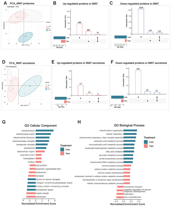
Exercise training and cold exposure both improve systemic metabolism, but the mechanisms are not well-established. We tested the hypothesis that adaptations to inguinal white adipose tissue (iWAT) are critical for these beneficial effects by determining the impact of exercise-trained and cold-exposed iWAT on systemic glucose metabolism and the iWAT proteome and secretome. Transplanting trained iWAT into sedentary mice improved glucose tolerance, while cold-exposed iWAT transplantation showed no such benefit. Compared to training, cold led to more pronounced alterations in the iWAT proteome and secretome, downregulating >2,000 proteins but also boosting iWAT's thermogenic capacity. In contrast, only training increased extracellular space and vesicle transport proteins, and only training upregulated proteins that correlate with favorable fasting glucose, suggesting fundamental changes in trained iWAT that mediate tissue-to-tissue communication. This study defines the unique exercise training- and cold exposure-induced iWAT proteomes, revealing distinct mechanisms for the beneficial effects of these interventions on metabolic health.

Keywords: adipose tissue; cold; exercise; proteomics; secretome; transplantation.


Conflict of interest statement

DECLARATION OF INTERESTS The authors declare no competing interests. R.J.W.M. and L.J.G. have received research support from Novo Nordisk, which is unrelated to this work.

Figures

Figure 1
Figure 1. iWAT transplantation from exercise trained but not cold-exposed mice improved glucose tolerance.
(A) iWAT was collected from male mice after 11 days of exercise, cold exposure, or a sedentary lifestyle and transplanted into male C57BL6 recipient mice. All analyses were conducted nine days post-transplantation. (B) Intraperitoneal glucose tolerance test (ipGTT) and area under the curve (AUC) in recipient mice after 9 days from transplantation. Data are presented as mean±SEM and were compared using One-way ANOVA. *p<0.05,**p<0.01, and ***p<0.001
Figure 2
Figure 2. Phenotypic responses of mice and their iWATs to exercise training and cold exposure
(A) Study design to perform quantitative proteomics of whole iWAT and secretome proteins from sedentary, exercise trained and cold-exposed mice. (B-D) Change in body weight (B), food intake (C) and glucose after 6hrs of fasting (D) of sedentary (gray), exercise trained (pink) and cold-exposed (green) mice (n=12/group). (E) H&E-stained section of iWAT from sedentary, trained and cold-exposed mice. Scale bar 1000μm (4x, up) and 50 μM (20x, down). (F) Adipocyte cell count per field measurement of iWAT from sedentary (gray), exercise trained (pink) and cold-exposed (green) mice (n=5/group). (G-I) Protein concentration (G), iWAT mass weight (H) and total protein/tissue weight ratio (I) for iWAT from sedentary, exercise trained and cold-exposed mice used for tissue proteomics analysis (n=3-4/group). Data are presented as mean±SEM and were compared using One-way ANOVA. *p<0.05,**p<0.01, and ***p<0.001
Figure 3
Figure 3. Distinct proteome and secretomic profiles of the exercise and cold-exposed iWATs
(A) PCA plot, including all protein quantified in iWAT from sedentary, exercise trained and cold-exposed mice (n=4 sedentary, 4 training, 3 cold). (B-C) UpSet intersection diagram showing the upregulated (B) and down-regulated (C) proteins compared to sedentary in iWAT from exercise trained (pink) and cold-exposed (green) mice. (D) PCA plot, including all protein quantified in iWAT secretome from sedentary, exercise trained and cold-exposed mice (n=3 sedentary, 4 training, 4 cold). (E-F) UpSet intersection diagram showing the upregulated (E) and down-regulated (F) proteins compared to sedentary in iWAT secretome from exercise trained (pink) and cold-exposed (green) mice. (G) Results of Gene Set Enrichment (GSE) analysis for cellular components of proteins that significantly change in iWAT following exercise training (pink) and cold exposure (green). The pathways shown here are considered significant after false discovery correction p < 0.05. (H) Results of Gene Set Enrichment (GSE) analysis for biological process of proteins that significantly change in iWAT following exercise training (pink) and cold exposure (green). The pathways shown here are considered significant after false discovery correction p < 0.05.
Figure 4
Figure 4. Top 50 differentially expressed proteins in iWAT: Insights into glucose metabolism and Rilpl2’s role in exercise-induced molecular responses
(A) Global correlation matrix of fasting glucose and the top 50 proteins changing in iWAT with exercise training (left) and cold exposure (right). The color of the circles in the matrix represents the level of correlation for the values that reach statistical significance (P<0.05); blue represents positive correlation and red represents negative correlation. (B) Representative images with relative quantification of Rilpl2 protein detected by western blot in iWAT from sedentary (gray), exercise training (pink) and cold-exposed (green) mice (n=3-4/group). (C ) Protein abundance level for Rilpl2 detected in the proteome dataset of iWAT from sedentary (gray), exercise training (pink) and cold-exposed (green) mice (n=3-4/group). (D) Protein-protein interaction network analysis of Rilpl2 using STRING database with the highest interaction confidence score (0.9) Data are presented as mean±SEM and were compared using One-way ANOVA. *p<0.05, and ***p<0.001
Figure 5
Figure 5. Expression patterns of the Rilpl2 protein network in iWAT in response to exercise training
(A-C) Visium images (A-B) and relative individual violin plots (C) showing the Rilpl2 expression level across the cell clusters detected in iWAT from sedentary (left) and exercise (right). (D-F) Individual violin plots showing the expression levels and distribution of Myo5a (D), Rab8a (E) and Rab10 (F) across cell clusters detected in iWAT from sedentary (left) and exercise (right).
Figure 6
Figure 6. Adipocyte-derived extracellular vesicles secretion in response to exercise training
(A) Study design to isolate Adipocyte-derived extracellular vesicles (Ad-EVs) from fresh isolated mature adipocytes from iWAT of sedentary and trained mice, using the size exclusion chromatography (SEC). (B) Representative electron micrograph showing CD63 gold immunostaining (1:20, 10nm) of Ad-EVs isolated from mature adipocytes using SEC. Scale bar: 100nM. (C-D) Nanoparticle Tracking Analysis (NTA) of Ad-EVs isolated from sedentary and trained iWAT (n=3 biological replicates, from two Ad-EVs isolation) representing the size (C) in nm and the number of particles per mL (C) of adipocyte suspension. Data are presented as mean±SEM and were compared using unpaired two-tailed Student’s t-test. **p<0.01.

References

    1. Murlasits Z., Kupai K., and Kneffel Z. (2022). Role of physical activity and cardiorespiratory fitness in metabolically healthy obesity: a narrative review. BMJ Open Sport Exerc Med 8, e001458. - PMC - PubMed
    1. Thyfault J.P., and Bergouignan A. (2020). Exercise and metabolic health: beyond skeletal muscle. Diabetologia 63, 1464–1474. - PMC - PubMed
    1. Stanford K.I., Middelbeek R.J.W., Townsend K.L., Lee M.-Y., Takahashi H., So K., Hitchcox K.M., Markan K.R., Hellbach K., Hirshman M.F., et al. (2015). A novel role for subcutaneous adipose tissue in exercise-induced improvements in glucose homeostasis. Diabetes 64, 2002–2014. - PMC - PubMed
    1. Nigro P., Vamvini M., Yang J., Caputo T., Ho L.-L., Carbone N.P., Papadopoulos D., Conlin R., He J., Hirshman M.F., et al. (2023). Exercise training remodels inguinal white adipose tissue through adaptations in innervation, vascularization, and the extracellular matrix. Cell Rep. 42, 112392. - PMC - PubMed
    1. Nigro P., Middelbeek R.J.W., Alves C.R.R., Rovira-Llopis S., Ramachandran K., Rowland L.A., Møller A.B., Takahashi H., Alves-Wagner A.B., Vamvini M., et al. (2021). Exercise Training Promotes Sex-Specific Adaptations in Mouse Inguinal White Adipose Tissue. Diabetes 70, 1250–1264. - PMC - PubMed

Publication types