

This is a preprint.
It has not yet been peer reviewed by a journal.
The National Library of Medicine is
running a pilot
to include preprints that result from research funded by NIH in PMC and PubMed.
[Preprint]. 2023 Oct 26:2023.10.16.562593.
doi: 10.1101/2023.10.16.562593.
Functional Tissue Units in the Human Reference Atlas
Affiliations
- PMID: 37905079
- PMCID: PMC10614912
- DOI: 10.1101/2023.10.16.562593
Item in Clipboard
Functional Tissue Units in the Human Reference Atlas
bioRxiv.
.
Update in
-
Functional tissue units in the Human Reference Atlas.Nat Commun. 2025 Feb 11;16(1):1526. doi: 10.1038/s41467-024-54591-6. Nat Commun. 2025. PMID: 39934102 Free PMC article.
Abstract
Functional tissue units (FTUs) form the basic building blocks of organs and are important for understanding and modeling the healthy physiological function of the organ and changes during disease states. In this first comprehensive catalog of FTUs, we document the definition, physical dimensions, vasculature, and cellular composition of 22 anatomically correct, nested functional tissue units (FTUs) in 10 healthy human organs. The catalog includes datasets, illustrations, an interactive online FTU explorer, and a large printable poster. All data and code are freely available. This is part of a larger ongoing international effort to construct a Human Reference Atlas (HRA) of all cells in the human body.
Figures
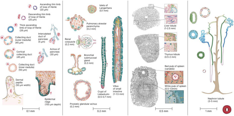
2D illustrations of all 22 FTU with name and size annotations.

HRA Interactive Functional Tissue Unit Explorer showing the 2D illustration of the renal corpuscle of the kidney along with associated experimental data.
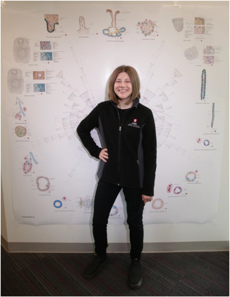
Poster of the HRA illustrating radial tree graphs of (1) the nested partonomy of organ anatomical structures and cell types of the human body with an overlay of (2) the branching structure of the blood vasculature extending from the heart (center of the figure) to FTUs (outer edge). Illustrations of all 22 FTUs are placed outside of the radial tree visualization for easy reference. One of the authors is posing for a photo.
References
-
- Osumi-Sutherland D. et al. Cell type ontologies of the Human Cell Atlas. Nat. Cell Biol. 23, 1129–1135 (2021). - PubMed
-
- Nephron ∣ Definition, Function, Structure, Diagram, & Facts ∣ Britannica. https://www.britannica.com/science/nephron (2023).
Publication types
Grants and funding
LinkOut - more resources
Full Text Sources