[Preprint]. 2023 Oct 19:2023.10.17.562769.
doi: 10.1101/2023.10.17.562769.

Nanomechanics of wild-type and mutant dimers of the tip-link protein protocadherin 15

Affiliations

Nanomechanics of wild-type and mutant dimers of the tip-link protein protocadherin 15

Camila M Villasante et al. bioRxiv. .

Update in

Abstract

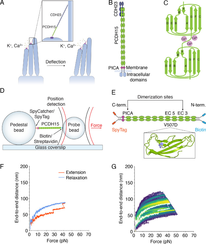
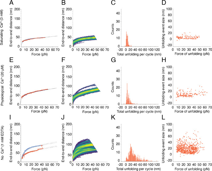
Mechanical force controls the opening and closing of mechanosensitive ion channels atop the hair bundles of the inner ear. The filamentous tip link connecting transduction channels to the tallest neighboring stereocilium modulates the force transmitted to the channels and thus changes their probability of opening. Each tip link comprises four molecules: a dimer of protocadherin 15 and a dimer of cadherin 23, all of which are stabilized by Ca2+ binding. Using a high-speed optical trap to examine dimeric PCDH15, we find that the protein's configuration is sensitive to Ca2+ and that the molecule exhibits limited unfolding at a physiological Ca2+ concentration. PCDH15 can therefore modulate its stiffness without undergoing large unfolding events in physiological Ca2+ conditions. The experimentally determined stiffness of PCDH15 accords with published values for the stiffness of the gating spring, the mechanical element that controls the opening of mechanotransduction channels. When PCDH15 has a point mutation, V507D, associated with non-syndromic hearing loss, unfolding events occur more frequently under tension and refolding events occur less often than in the wild-type protein. Our results suggest that the maintenance of appropriate tension in the gating spring is critical to the appropriate transmission of force to transduction channels, and hence to hearing.


Figures

Figure 1.
Figure 1.. The structure of PCDH15 and measurements with an optical trap.
(A) When stereocilia are deflected towards the tall edge of a hair bundle, the tip links (inset) connecting them stretch, opening the mechanotransduction channels (purple) atop each stereocilium and allowing the ions within the endolymph to flow into the cell, resulting in depolarization. (B) PCDH15 comprises 11 EC domains and a PICA domain at its carboxy terminus. PCDH15 binds with CDH23 at its amino terminus in a handshake interaction. (C) EC domains are composed primarily of β-sheets. Ca2+ binding in the linker regions and at the edges of the domains stabilizes the structure against unfolding. (D) In our apparatus, the protein is tethered between two beads and two laser beams act on the probe bead to measure its position and exert force on it. (E) In our experimental construct, we maintained the dimerization sites in PCDH15 while adding two disulfide bonds to each end in addition to the distinct molecular tags at each end. The V507D construct was identical to the wild-type construct save for the insertion of the mutation in place of V507 (purple in inset). (F) A force-ramp experiment comprises the extension phase of the cycle, during which force is increased at a constant rate, and the relaxation phase of the cycle, during which force is decreased back to a minimum at the same constant rate. In these experiments, the minimum force is 1 pN and 2 s elapse between successive cycles. Unfolding events can be seen as sudden steps during an individual extension. (G) Repetition of a force-ramp cycle hundreds of times on the same protein molecule yields a heat map in which the brighter colors represent more highly occupied states. The heatmap superimposes both extension and relaxation phases of every cycle. The illustrative cycle from panel F is overlaid in white.
Figure 2.
Figure 2.. Force-ramp responses of wild-type PCDH15 at three Ca2+ concentrations.
(A) At a saturating Ca2+ concentration of 3 mM, PCDH15 unfolds infrequently. The dashed line represents a fit of our model, Equation (1), to the extension and relaxation phases of the illustrative cycle. (B) In a heatmap for a saturating level of Ca2+, 3 mM, the single bright branch indicates one highly occupied state that reflects the infrequent and small unfolding events. (C) The total unfolding length during the extension phases of all cycles in all datasets at 3 mM Ca2+ peaked at 1.8 ± 0.1 nm and 6.3 ± 0.2 nm (means ± SEMs; N = 5 datasets; n = 131 events). (D) Many unfolding events occurred at forces below 10 pN at 3 mM Ca2+. (E) At a physiological Ca2+ concentration of 20 μM, PCDH15 often does not unfold and is more extensible than under 3 mM Ca2+ in the same force range. (F) The illustrative heatmap has one bright branch indicative of the infrequent and small unfolding events at 20 μM Ca2+. (G) At 20 μM Ca2+, the total unfolding per cycle was slightly greater than at a saturating level of Ca2+, with peaks at 4.5 ± 0.1 nm and 11.5 ± 0.1 nm (means ± SEMs; N = 4 datasets; n = 172 events). (H) At 20 μM Ca2+, individual unfolding events with a mean of 4.6 ± 0.1 nm occurred predominantly at forces below 40 pN. (I) An illustrative force-ramp cycle in the absence of Ca2+ and in the presence of 1 mM EDTA shows a small unfolding event followed by a larger one. In this case, our protein model, Equation (1), is fitted separately to each segment demarcated by the unfolding events. (J) The numerous bright branches in the heatmap reflect the multiple preferred conformational states of PCDH15 in the absence of Ca2+ and in the presence of 1 mM EDTA. (K) The total unfolding per cycle peaked at 23.3 ± 0.6 nm (mean ± SEM; N = 6 datasets, n = 490 events), but many larger events occurred in the absence of Ca2+ and in the presence of 1 mM EDTA. (L) There is no clear relationship between the size of individual unfolding events and the corresponding forces in the absence of Ca2+ and in the presence of 1 mM EDTA.
Figure 3.
Figure 3.. Force-ramp responses of V507D at three Ca2+ concentrations.
(A) At a saturating level of Ca2+, 3 mM, a subset of V507D molecules underwent only small unfolding events while another subset had more frequent unfolding (Figure S5A). The dashed line represents the fit of our model, Equation (1), to the segmented extension and relaxation phases. (B) The bright branches on the heatmap reflect the frequent unfolding seen in the individual cycle. (C) The frequency distribution of total unfolding length per cycle peaked around 8.8 ± 0.1 nm, 50.0 ± 0.2 nm, and 66.3 ± 0.1 nm (means ± SEMs; N = 24 datasets; n = 1889 events). (D) As the size of the individual unfolding event got larger, the force of unfolding tended to be lower, with many events occurring around 30 pN. (E) At a physiological concentration of Ca2+, 20 μM, frequent unfolding was seen on the single cycle level. (F) The unfolding seen in the individual cycles is reflected by the bright branches on the exemplary heatmap. (G) The frequency distribution of total unfolding length per cycle during the extension phase peaked around 14.2 ± 0.2 nm, 27.3 ± 0.1 nm, and 39.4 ± 0.3 nm (means ± SEMs; N = 16 datasets; n = 1081 events). (H) As at a saturating level of Ca2+, the larger unfolding events were associated with smaller forces of unfolding. (I) When Ca2+ was absent, V507D extended easily, even without large unfolding events. (J) The bright branches on the illustrative heatmap reflect the unfolding behavior seen on the individual cycle level. The upper limits of the end-to-end distance at this level of Ca2+ exceeded those seen at higher concentrations of Ca2+. (K) The frequency distribution of total unfolding length per cycle was unimodal, with one peak at 49.3 ± 1.4 nm (mean ± SEM; N = 4 datasets; n = 362 events). (L) In the absence of Ca2+, there was no clear relationship between the size of individual unfolding events and the forces at which they occurred.
Figure 4.
Figure 4.. Clustering of force-ramp trajectories and inter-state transitions.
(A) In a heatmap representing the relaxation-phase trajectories from all Ca2+ conditions and both PCDH15 constructs the lines with different colors demarcate the six conformational states. (B) The fitted xE values for individual trajectories at different conformational states are shown in the scatter plot. The data represent means ± SEMs. (C-H) Conformational transition maps summarize the trajectories of the two PCDH15 constructs at different Ca2+ concentrations. Segmented by conformational changes, the values along the abscissa denote individual segment within a trajectory in ascending order of force. The thickness of each arrow shaft denotes the frequency of a specific state transition. The horizontal arrows indicate conformational changes within a state, whereas the arrows pointing up and to the right denote conformational changes made between distinct states.
Figure 5.
Figure 5.. Entropic elasticity of PCDH15’s states as a function of force.
The stiffnesses of the states were calculated by finding the inverse of the mean slope of each state. In state 1, when PCDH15 is fully folded, the stiffness approached the enthalpic values above 15 pN. In states 2–6, in which PCDH15 had some unfolded portions, the stiffness of PCDH15 remained below the enthalpic limit. State 1 largely comprises trajectories of the wild-type protein at a saturating level of Ca2+, whereas the remaining states predominantly contain trajectories at lower Ca2+ concentrations and from the V507D mutant. The results suggest that, under physiological conditions, the effective stiffness of PCDH15 is lower than its enthalpic limit of 40 mN×m−1 at a saturating concentration of Ca2+. Error bars represent SEMs.
Figure 6.
Figure 6.. Proposed mechanism of deafness for mutated V507D.
(A) Our data suggest that V507D is misfolded or partially unfolded over the physiological range of forces experienced in the cochlea. (B) If the tip links lack sufficient tension to open the mechanosensitive ion channels when stimulated, the ensuing deficit of Ca2+ in the stereocilia would cause their degeneration.

References

    1. Gillespie P. G. & Müller U. Mechanotransduction by Hair Cells: Models, Molecules, and Mechanisms. Cell 139, 33–44 (2009). - PMC - PubMed
    1. Kandel Eric, Koester John & Mack S. Principles of Neural Science. (McGraw Hill/Medical, 2021).
    1. Kazmierczak P. et al. Cadherin 23 and protocadherin 15 interact to form tip-link filaments in sensory hair cells. Nature 449, 87–91 (2007). - PubMed
    1. Assad J. A., Shepherd G. M. & Corey D. P. Tip-link integrity and mechanical transduction in vertebrate hair cells. Neuron 7, 985–994 (1991). - PubMed
    1. Zhao Y.-d., Yamoah E. N. & Gillespie P. G. Regeneration of broken tip links and restoration of mechanical transduction in hair cells. Proc. Natl. Acad. Sci. 93, 15469–15474 (1996). - PMC - PubMed

Publication types