[Preprint]. 2023 Oct 17:2023.10.15.562341.
doi: 10.1101/2023.10.15.562341.

hkb is required for DIP-α expression and target recognition in the Drosophila neuromuscular circuit

Affiliations

hkb is required for DIP-α expression and target recognition in the Drosophila neuromuscular circuit

Yupu Wang et al. bioRxiv. .

Update in

Abstract

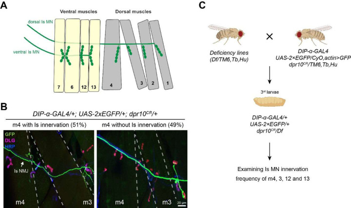
Our nervous system contains billions of neurons that form precise connections with each other through interactions between cell surface proteins (CSPs). In Drosophila, the Dpr and DIP immunoglobulin protein subfamilies form homophilic or heterophilic interactions to instruct synaptic connectivity, synaptic growth and cell survival. However, the upstream regulation and downstream signaling mechanisms of Dprs and DIPs are not clear. In the Drosophila larval neuromuscular system, DIP-α is expressed in the dorsal and ventral type-Is motor neurons (MNs). We conducted an F1 dominant modifier genetic screen to identify regulators of Dprs and DIPs. We found that the transcription factor, huckebein (hkb), genetically interacts with DIP-α and is important for target recognition specifically in the dorsal Is MN, but not the ventral Is MN. Loss of hkb led to complete removal of DIP-α expression. We then confirmed that this specificity is through the dorsal Is MN specific transcription factor, even-skipped (eve), which acts downstream of hkb. Genetic interaction between hkb and eve revealed that they act in the same pathway to regulate dorsal Is MN connectivity. Our study provides insight into the transcriptional regulation of DIP-α and suggests that distinct regulatory mechanisms exist for the same CSP in different neurons.


Figures

Figure 1.
Figure 1.
Establishing a sensitized background for deficiency screen. A. Cartoon depicting the innervation pattern of dorsal Is MN and ventral Is MN. B. Representative images of muscle 4 with Is innervation or without Is innervation, in trans-heterozygotes of DIP-α (the DIP-α-GAL4 is also a null allele) and dpr10. 51% of m4 are innervated by the dorsal Is MN. GFP (green), DLG (magenta) and HRP (blue) are shown in the images. Arrow pointing to Is NMJ. C. Workflow of deficiency screen. Male flies carrying deficiency chromosome were crossed to females with DIP-α and dpr10 mutations. Female third instar larvae were selected against the second and third chromosome balancers (CyO,actin>GFP and TM6,Tb,Hu). Triple-heterozygous larvae were dissected and Is innervation frequency on m4, 3, 12 and 13 were scored.
Figure 2.
Figure 2.
Deficiency screen revealed candidate regions that cover genetic interactors of DIP-α or dpr10. A-D. Is innervation frequency on (A) m4, (B) m3, (C) m12 and (D) m13. The red column indicates the control innervation frequency from the sensitized background (trans-heterozygotes of DIP-α and dpr10). Grey columns are non-significant from control whereas the yellow columns are the deficiency lines that show significantly different innervation frequencies compared to control. The cut-off p-values are indicated by dashed lines. Asterisk indicates ED5100.
Figure 3.
Figure 3.
A sub-screen identified huckebein (hkb) as a genetic interactor. A. Cartoon depicting the deleted regions within deficiency lines ED5100, ED5142, and ED5046. B. Quantification shows a significant reduction of m4-Is innervation when combining ED5046, but not ED5142, with the sensitized background, suggesting the shared region between the original deficiency line (ED5100) and ED5046 covers the candidate gene(s). N (NMJs) = 177, 196, 155 and 155. p-values are indicted. C. Quantification of sub-screen of individual genes from candidate region shown in (A). Alleles used to create triple-heterozygotes are, auxD128, abs00620, cpxMI00784, vps24EY04708, hkb2, contG5080, tub2, lostEY11645. Note that huckebein (hkb2) further reduced m4-Is innervation frequency. N (NMJs) = 177, 138, 62, 58, 29, 135, 39, 77, 78 and 57. p-values are indicted.
Figure 4.
Figure 4.
hkb genetically interacts with DIP-α, but not dpr10. A. Genetic interaction assay between hkb and dpr10. Single heterozygotes of dpr10 or hkb did not have altered m4-Is innervation, and neither did the trans-heterozygotes. N (NMJs) = 93, 154, 83, 125, 102 and 149. p-values are indicted. B. Genetic interaction assay between hkb and DIP-α. Single heterozygotes of DIP-α or hkb had slightly decreased m4-Is innervation frequency, while the trans-heterozygotes showed a further reduction, suggesting that hkb genetically interacts with DIP-α. N (NMJs) = 114, 105, 106, 103, 99 and 103. p-values are indicted.
Figure 5.
Figure 5.
hkb is required for DIP-α-EGFP expression in dorsal Is MNs. A. Cartoon depicting the focal plane in B-D. B and B’. Representative images of dorsal Is MN cell bodies (arrows in B) and ventral Is MN cell bodies (arrows in B’) labeled with GFP (green), Eve (red) and FasII (magenta), in control embryos. DIP-α-EGFP is expressed in both dorsal and ventral Is MNs. C and C’. Representative images of dorsal Is MN cell bodies (dash circles in C) and ventral Is MN cell bodies (arrows in C’) in hkb2 mutant embryos. Eve and DIP-α-EGFP are missing in dorsal Is MNs, whereas DIP-α-EGFP expression in ventral Is MNs is not affected. D and D’. Representative images of dorsal Is MN cell bodies (dash circles in D) and ventral Is MN cell bodies (arrows in D’) in heteroallelic hkb mutant embryos (hkb2/hkbA321R1). Eve and DIP-α-EGFP are missing in dorsal Is MNs, whereas DIP-α-EGFP expression in ventral Is MNs is not affected.
Figure 6.
Figure 6.
hkb functions through eve to regulate DIP-α-EGFP expression. A and A’. Representative images of dorsal Is MN cell bodies (arrows in A) and ventral Is MN cell bodies (arrows in A’) labeled with GFP (green), Eve (red) and FasII (magenta), in control embryos. DIP-α-EGFP is expressed in both dorsal and ventral Is MNs. B and B’. Representative images of dorsal Is MN cell bodies (dash circles in B) and ventral Is MN cell bodies (arrows in B’) in eveΔRN2 mutant embryos. Eve and DIP-α-EGFP are missing in dorsal Is MNs, whereas DIP-α-EGFP expression in ventral Is MNs is not affected. C. Genetic interaction assay between eve and DIP-α. Single heterozygotes of eve or DIP-α did not have altered m4-Is innervation, while the trans-heterozygotes showed a further reduction, suggesting that eve genetically interacts with DIP-α. N (NMJs) = 102, 104, 84 and 104. p-values are indicted. D. Genetic interaction assay between hkb and eve. Single heterozygotes of hkb or eve did not have altered m4-Is innervation, while the trans-heterozygotes showed a further reduction, suggesting that hkb genetically interacts with eve to regulate Is innervation. N (NMJs) = 103, 87, 105, 81, 112 and 86. p-values are indicted.

References

    1. Ashley J., Sorrentino V., Lobb-Rabe M., Nagarkar-Jaiswal S., Tan L., Xu S., Xiao Q., Zinn K. and Carrillo R. A. (2019). Transsynaptic interactions between IgSF proteins DIP-α and Dpr10 are required for motor neuron targeting specificity. eLife 8, e42690. - PMC - PubMed
    1. Barish S., Nuss S., Strunilin I., Bao S., Mukherjee S., Jones C. D. and Volkan P. C. (2018). Combinations of DIPs and Dprs control organization of olfactory receptor neuron terminals in Drosophila. Plos Genet 14, e1007560. - PMC - PubMed
    1. Bornstein B., Meltzer H., Adler R., Alyagor I., Berkun V., Cummings G., Reh F., Keren-Shaul H., David E., Riemensperger T., et al. (2021). Transneuronal Dpr12/DIP-δ interactions facilitate compartmentalized dopaminergic innervation of Drosophila mushroom body axons. Embo J e105763. - PMC - PubMed
    1. Bossing T., Technau G. M. and Doe C. Q. (1996). huckebein is required for glial development and axon pathfinding in the neuroblast 1–1 and neuroblast 2–2 lineages in the Drosophila central nervous system. Mech Develop 55, 53–64. - PubMed
    1. Broadus J., Skeath J. B., Spana E. P., Bossing T., Technau G. and Doe C. Q. (1995). New neuroblast markers and the origin of the aCC/pCC neurons in the Drosophila central nervous system. Mech Develop 53, 393–402. - PubMed

Publication types