Case Reports
. 2023 Sep 28;15(9):e46167.
doi: 10.7759/cureus.46167. eCollection 2023 Sep.

Cemento-Ossifying Fibroid Epulis in the Posterior Maxilla

Affiliations
Case Reports

Cemento-Ossifying Fibroid Epulis in the Posterior Maxilla

Vijaya Lakshmi G et al. Cureus. .

Abstract

Cemento-ossifying fibroma is a benign fibro-osseous lesion arising from the periodontal ligament and has the potential to form cementum and bone in the periodontal ligament. Cemento-ossifying fibroma is a painless, pedunculated, or sessile, smooth exophytic growth arising attached to the gingival tissues. We present a case of cemento-ossifying fibroid epulis in the posterior maxilla attached to the interdental gingiva between the 26 and 27 region buccally in a 52-year-old female patient managed with surgical excision of the lesion, extraction of the involved teeth, curettage, and palatal obturator while under general anesthesia. The patient was followed up post-operatively, healing was satisfactory, there were no signs of infection, and no recurrence was noted in the six-month follow-up period.

Keywords: calcifications; cemento-ossifying fibroma; fibroma; gingiva; ossifying; periodontal ligament.


Conflict of interest statement

The authors have declared that no competing interests exist.

Figures

Figure 1
Figure 1. Pre-op clinical image
3x2 cm solitary swelling was noted in relation to 26, 27 region buccally, which is oval in shape, pedunculated attached to the interdental gingiva between 26 and 27 region and pink in color and has smooth texture.
Figure 2
Figure 2. Pre-operative OPG
OPG reveals a radiopaque rim surrounding the radiolucent region superimposed over the 26, 27, and 28 region. OPG: Orthopantomogram
Figure 3
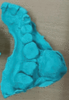
Figure 3. Model of the lesion
The maxillary cast shows a pedunculated lesion attached to the interdental gingiva between the 26 and 27 region.
Figure 4
Figure 4. Model of the lesion
The maxillary cast on a lateral aspect shows that a 3x2 cm solitary swelling was noted in the 26 and 27 region buccally.
Figure 5
Figure 5. Excision of the lesion
Surgical excision of the lesion was performed along with the pedicle and the interdental gingiva.
Figure 6
Figure 6. Osteoplasty post excision
Osteoplasty was performed after excision and bone was filed.
Figure 7
Figure 7. Excised specimen
Excised lesion along with the pedicle.
Figure 8
Figure 8. Palatal obturator in situ
Coe-pack was placed in the operated site, the prepared palatal obturator was adapted and secured to the teeth using 26-gauge stainless steel wires.
Figure 9
Figure 9. Histopathology is suggestive of cemento-ossifying fibroid epulis (fibroepithelial polyp with calcification)
The fibrous connective tissue shows dense interlacing bundles with fibroblasts and vascular spaces. Bony trabecular and cementum-like mass seen with the center of the lesion.
Figure 10
Figure 10. Histopathology is suggestive of cemento-ossifying fibroid epulis (fibroepithelial polyp with calcification)
Parakeratinized stratified squamous epithelium with long and slender rete ridges. Fibrocellular connective tissue with calcifications.
Figure 11
Figure 11. Post-operative image after excision
Post-op image shows satisfactory wound healing.
Figure 12
Figure 12. Post-operative OPG
Post-operative OPG shows no recurrence in the operated site in the six-month follow-up.

References

    1. Peripheral cemento-ossifying fibroma: a case report with review of literature. Mishra AK, Maru R, Dhodapkar SV, Jaiswal G, Kumar R, Punjabi H. World J Clin Cases. 2013;1:128–133. - PMC - PubMed
    1. Peripheral cemento-ossifying fibroma: case series literature review. Verma E, Chakki AB, Nagaral SC, Ganji KK. Case Rep Dent. 2013;2013:930870. - PMC - PubMed
    1. Peripheral cemento-ossifying fibroma - a rare case report. Kaushik N, Srivastava N, Rana V, Suhane C. J Cancer Res Ther. 2022;18:0–6. - PubMed
    1. Peripheral ossifying fibroma. Mohiuddin K, Priya NS, Ravindra S, Murthy S. https://pubmed.ncbi.nlm.nih.gov/24174733/ J Indian Soc Periodontol. 2013;17:507–509. - PMC - PubMed
    1. Peripheral ossifying fibroma: a 20-year retrospective study with focus on clinical and morphological features. Cavalcante IL, Barros CC, Cruz VM, et al. Med Oral Patol Oral Cir Bucal. 2022;27:0–7. - PMC - PubMed

Publication types

LinkOut - more resources