

Metformin Inhibits the Estrogen-mediated Epithelial-Mesenchymal Transition of Ectopic Endometrial Stromal Cells in Endometriosis
- PMID: 37905623
- PMCID: PMC10621412
- DOI: 10.21873/invivo.13356
Metformin Inhibits the Estrogen-mediated Epithelial-Mesenchymal Transition of Ectopic Endometrial Stromal Cells in Endometriosis
Abstract
Background/aim: Endometriosis is an estrogen-dependent disease characterized by the ectopic implantation and growth of endometrial tissue outside the uterus. Endometrial stromal cells (ESCs) play a crucial role in the pathogenesis of endometriosis. Epithelial-mesenchymal transition (EMT) has recently been described in endometriosis and was induced by estrogen. Metformin has been shown to inhibit EMT in various diseases, but its role in endometriosis remains unclear.
Materials and methods: We collected endometrial tissue samples from patients with endometriosis and healthy controls and isolated primary ESCs. We performed gene expression analysis using the Gene Expression Omnibus (GEO) dataset and validated the results by immunohistochemistry in tissue samples. We also assessed the effects of metformin on the proliferation, migration and invasion of ectopic ESCs (EESCs) by Cell Counting Kit-8 and Transwell migration and invasion assays, respectively. We analyzed the protein expression of EMT-related markers (N-cadherin, vimentin, twist, and snail) and β-catenin by Western blotting and immunohistochemistry.
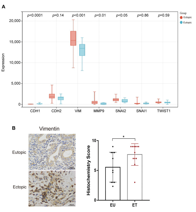
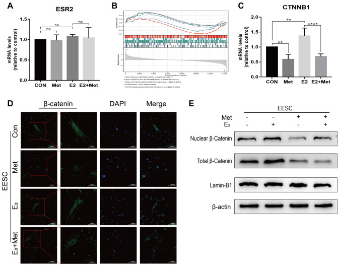
Results: We found that vimentin was highly expressed in ectopic endometrial tissues compared to normal endometrial tissues. Metformin treatment inhibited the proliferation, migration and invasion of EESCs in a dose-dependent manner. Metformin treatment also downregulated the expression of EMT-related markers and reduced the expression and nuclear translocation of β-catenin in EESCs.
Conclusion: Our results suggest that metformin inhibits estrogen-induced EMT and regulates the expression of β-catenin in EESCs. This study provides new insights into the potential therapeutic role of metformin in endometriosis.
Keywords: Endometriosis; endometrial stromal cells; epithelial-mesenchymal transition; estrogen; metformin; β-catenin.
Copyright © 2023, International Institute of Anticancer Research (Dr. George J. Delinasios), All rights reserved.
Conflict of interest statement
The Authors declare that they have no competing interests in relation to this study.
Figures

Similar articles
-
MicroRNA-126-5p downregulates BCAR3 expression to promote cell migration and invasion in endometriosis.Mol Cell Endocrinol. 2019 Aug 20;494:110486. doi: 10.1016/j.mce.2019.110486. Epub 2019 Jun 21. Mol Cell Endocrinol. 2019. PMID: 31233772
-
Hypoxia-inducible factor 1α-induced epithelial-mesenchymal transition of endometrial epithelial cells may contribute to the development of endometriosis.Hum Reprod. 2016 Jun;31(6):1327-38. doi: 10.1093/humrep/dew081. Epub 2016 Apr 19. Hum Reprod. 2016. PMID: 27094478
-
TIPE2 inhibits the migration and invasion of endometrial cells by targeting β-catenin to reverse epithelial-mesenchymal transition.Hum Reprod. 2020 Jun 1;35(6):1377-1390. doi: 10.1093/humrep/deaa062. Hum Reprod. 2020. PMID: 32469403
-
A review of the effects of estrogen and epithelial-mesenchymal transformation on intrauterine adhesion and endometriosis.Transpl Immunol. 2023 Aug;79:101679. doi: 10.1016/j.trim.2022.101679. Epub 2022 Jul 29. Transpl Immunol. 2023. PMID: 35908631 Review.
-
Estrogen- and Progesterone (P4)-Mediated Epigenetic Modifications of Endometrial Stromal Cells (EnSCs) and/or Mesenchymal Stem/Stromal Cells (MSCs) in the Etiopathogenesis of Endometriosis.Stem Cell Rev Rep. 2021 Aug;17(4):1174-1193. doi: 10.1007/s12015-020-10115-5. Epub 2021 Jan 7. Stem Cell Rev Rep. 2021. PMID: 33411206 Free PMC article. Review.
Cited by
-
Insights into the Molecular Mechanisms and Signaling Pathways of Epithelial to Mesenchymal Transition (EMT) in the Pathophysiology of Endometriosis.Int J Mol Sci. 2025 Aug 1;26(15):7460. doi: 10.3390/ijms26157460. Int J Mol Sci. 2025. PMID: 40806587 Free PMC article. Review.
References
MeSH terms
Substances
LinkOut - more resources
Full Text Sources
Medical
Research Materials